
Figure 2.
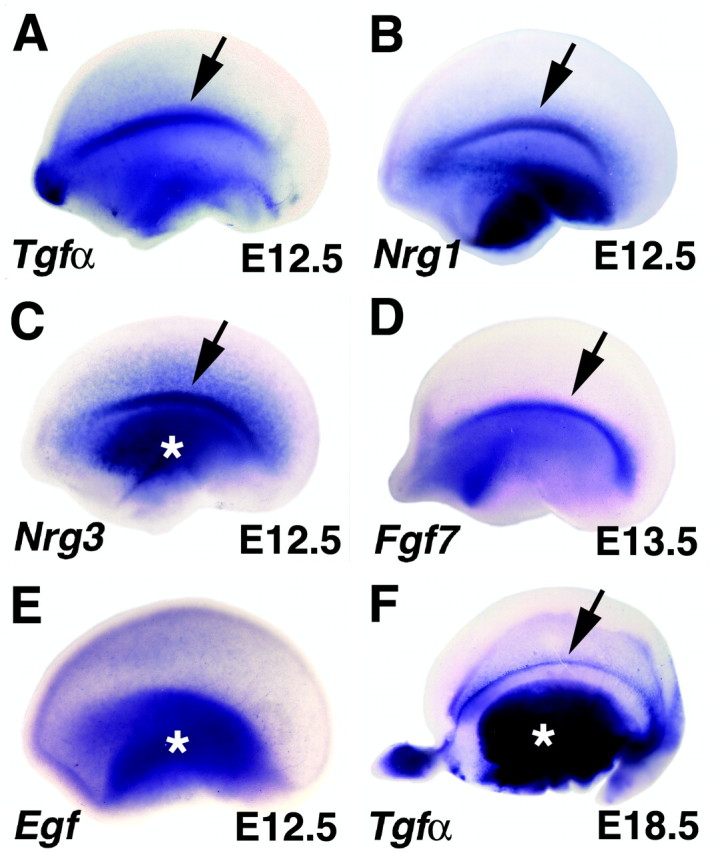
EGF family members are expressed in the cortical antihem. A-E, Embryonic cerebral hemispheres viewed from the lateral face; anterior to the left (A-C, E, E12.5; D, E13.5). F, E18.5 hemisphere viewed from the inside looking laterally. A-C, Peaks of expression of Tgfα, Nrg1, and Nrg3 mark the curving longitudinal lateral band of the antihem (arrows in A-C). Tgfα expression is maintained for several days in this position (arrow, F). D, Fgf7 is also expressed in the antihem at E13.5. E, The founding member of the EGF family, Egf itself, is expressed in the ventral telencephalon and in the cortical primordium without a peak of expression at the antihem. Asterisks mark expression in ventral telencephalon (C, E, F).