
Figure 4.
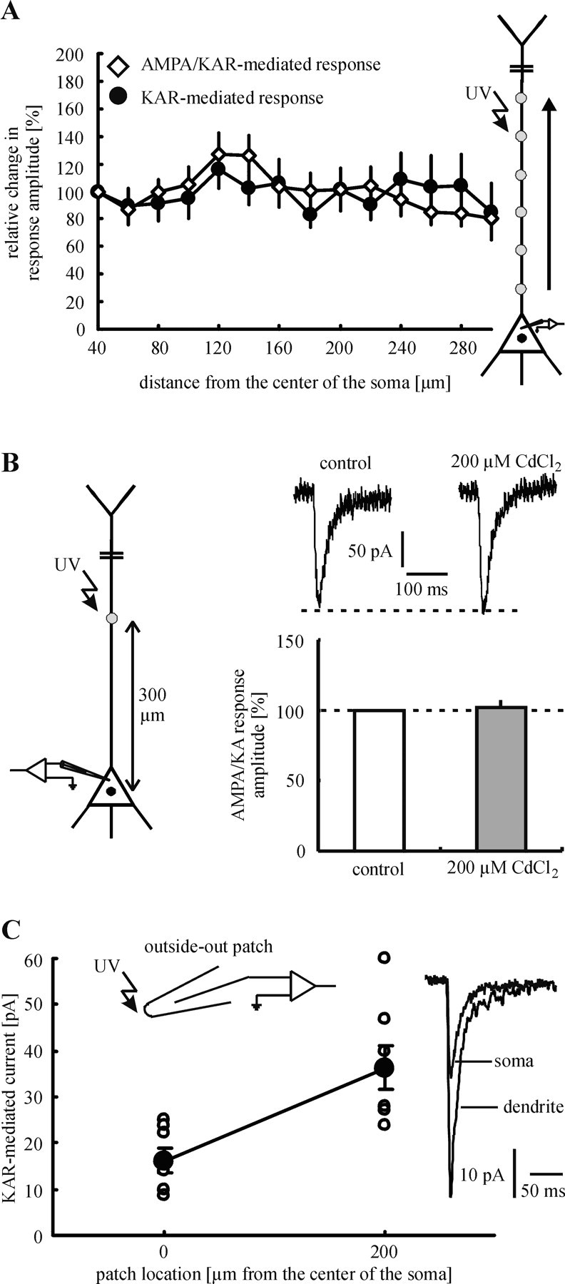
Subcellular distribution of KARs and AMPARs. A, Glutamate was applied photolytically to sites 20 μm apart along the apical dendrite. The profile of the dendritic glutamate sensitivity does not change after the addition of GYKI 53655. This result strongly suggests that the relative ratio of the number of KARs to AMPARs does not change toward the distal dendrite. B, Voltage-dependent Ca2+ channels do not affect the amplitude of mixed AMPA/KAR-mediated currents in our experiments. In the presence of d-AP-5 (control) mixed AMPA/KAR-mediated responses were evoked by glutamate application to the most distal part of the dendrite that was examined (300 μm from the center of the soma). To block voltage-dependent Ca2+ channels, we added CdCl2 (200 μm) to the superfusion medium. This pharmacological treatment changed neither the amplitude nor the size of the glutamate responses. C, Glutamate responses, which were evoked at somatic outside-out patches, showed smaller amplitudes on average than glutamate responses that were elicited at dendritic outside-out patches. Because of the evidence for identical unitary properties of somatic and dendritic KARs, this finding indicates an increasing density of KARs toward the distal dendrite.