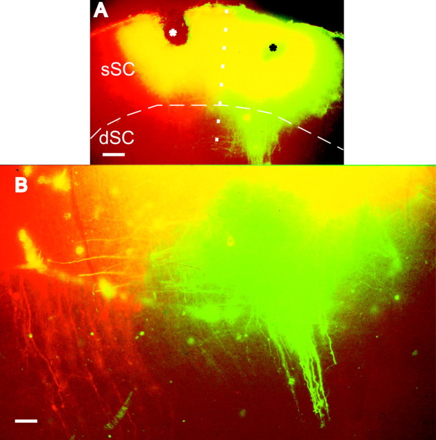
Figure 2.
Superficial- to deep-layer connections in infant ferrets. A, Low-power epifluorescent micrograph of coronal sections of the SC from a P20 ferret. Micrographs taken with the rhodamine filter have been digitally superimposed on images photographed using the fluorescein filter. Crystals of DiI and DiAsp were placed adjacent to each other in the mediolateral plane. Asterisks mark the location of the crystals in the superficial layers of the SC, and the dotted line indicates the midpoint between them. The dashed line indicates the presumptive border between sSC and dSC. B, High-power view of an adjacent section showing bundles of fibers emerging from the dye placement sites in the superficial SC and heading into the deeper layers perpendicular to the pial surface. Red and green fibers appear to descend separately, indicating that some topographic order is already present. Within the superficial layers, some fibers run parallel to the pial surface, appearing yellow in areas of overlap. Scale bars: A, 100 μm; B,50 μm.

