
Figure 9.
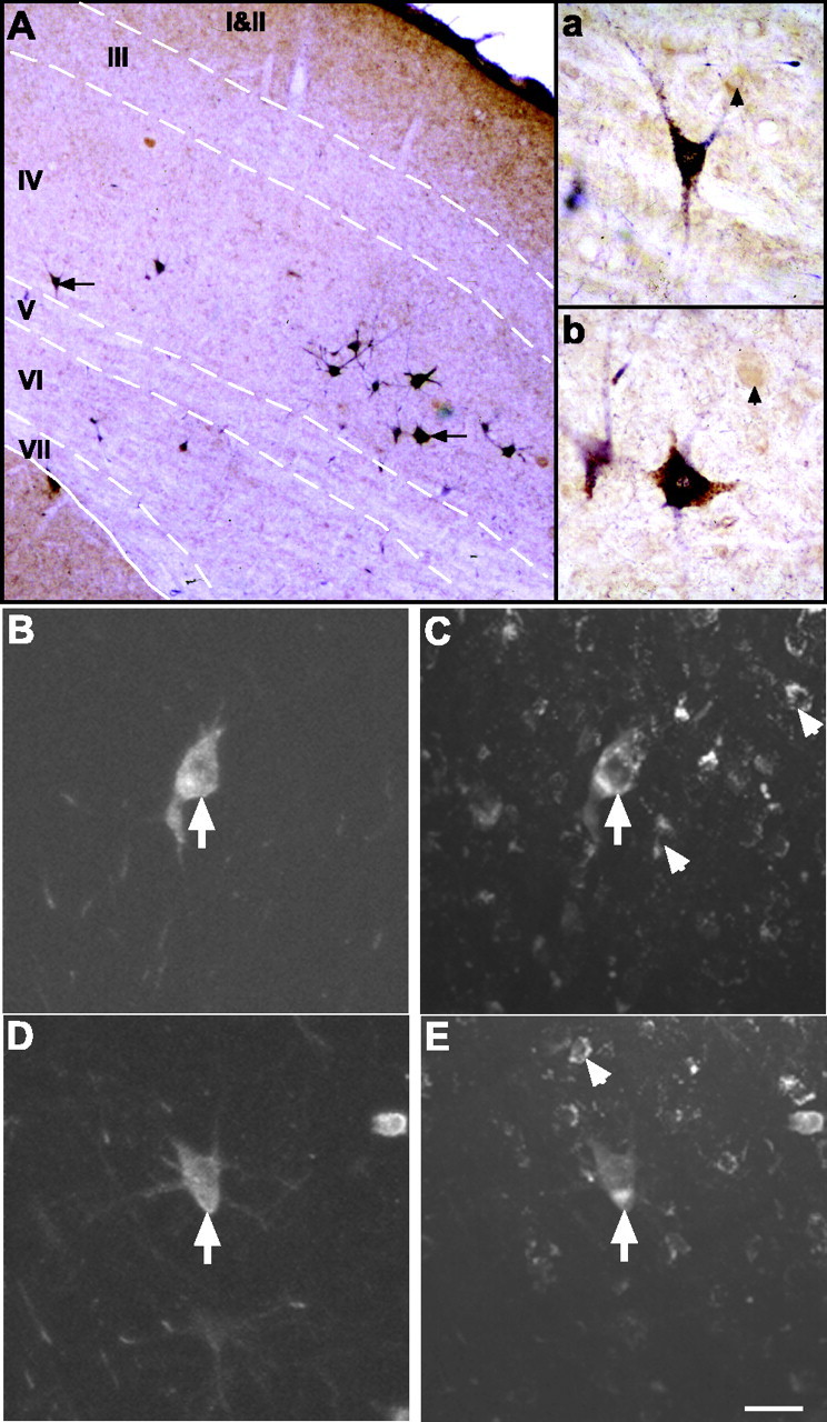
Double labeling of superior colliculus projection neurons. A, Injections of BDA into the tectoreticulospinal tract produced retrograde labeling in many medium and large multipolar neurons (arrows) located predominantly in layers IV and VI of the contralateral SC (dark-blue reaction product). The dashed lines indicate the layer borders. Double labeling with NR1 immunocytochemistry (brown reaction product) revealed that many of these tectoreticulospinal projection neurons expressed the NR1 subunit of the NMDA receptor. The two double-labeled cells indicated by the arrows are shown at higher power in the insets a and b. The arrowheads in a and b depict single-labeled NR1 cells. B-E, Confocal microscope images showing retrogradely labeled tectoreticulospinal neurons visualized with Cy5 (B and D) and, for the same fields of view, NR1 immunofluorescence visualized with Cy2 (E and F). Each image represents a projected maximum series of 16 focal planes spaced at 1 μm intervals. The BDA-labeled SC neurons marked by the arrows in B and D also show punctate, perinuclear NR1 immunofluorescence (arrows in C and E, respectively). The arrowheads in C and E indicate single-labeled NR1 cells. Scale bar: (in E) A, 75 μm; B-E, 20 μm.