
Figure 5.
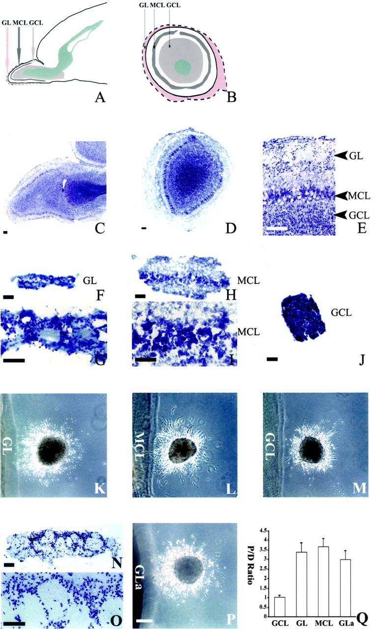
Dissection of OB layers with the chemoattractive activity. A, A diagram of the sagittal section of neonatal forebrain showing the anatomic layers of the OB. B, A diagram of the coronal section of the neonatal OB (outlined by dashed line). The layer between the GL and MCL is the external plexiform layer, whereas the layer between the MCL and GCL is the internal plexiform layer. C, A sagittal slice of the P4 OB stained with cresyl violet showing the different layers in the OB. D, A coronal slice of P4 OB stained with cresyl violet. E, A higher magnification view of C showing the GL, external plexiform layer, MCL, internal plexiform layer, and GCL. F, A GL explant dissected from a P4 OB was stained with cresyl violet. G, A higher magnification view of F showing that it contains GL. H, An explant dissected from a P4 OB was stained with cresyl violet, showing that it contains the external plexiform layer, MCL, and internal plexiform layer. I, A higher magnification view of H. J, A GCL explant from a P4 OB was stained with cresyl violet, showing that it contains only GCL but not the other layers in the OB. K, Effect of the GL explants on cells migrating from a P4 SVZa explant (n = 66 of 74). L, Effect of the MCL explants (including the MCL and parts of the external and internal plexiform layers) on cells migrating from a P4 SVZa explant (n = 58 of 61). M, Effect of GCL explants on cells migrating from a P4 SVZa explant (n = 69 of 77). N, A GL explant dissected from the adult OB was stained with cresyl violet, showing that it contains only the GL. O, A higher magnification view of N. P, Effect of an adult GL explant on cells migrating from a P4 SVZa explant (n = 87 of 93). GLa, Adult GL. Q, Effects of different layers from the OB on the distribution of cells migrating out of the SVZa explants. The cell numbers were counted from 24 P4 GCL explants, 26 P4 GL explants, 22 P4 MCL explants, and 25 adult GL (GLa) explants. Scale bars, 100 μm.