Figure 6.

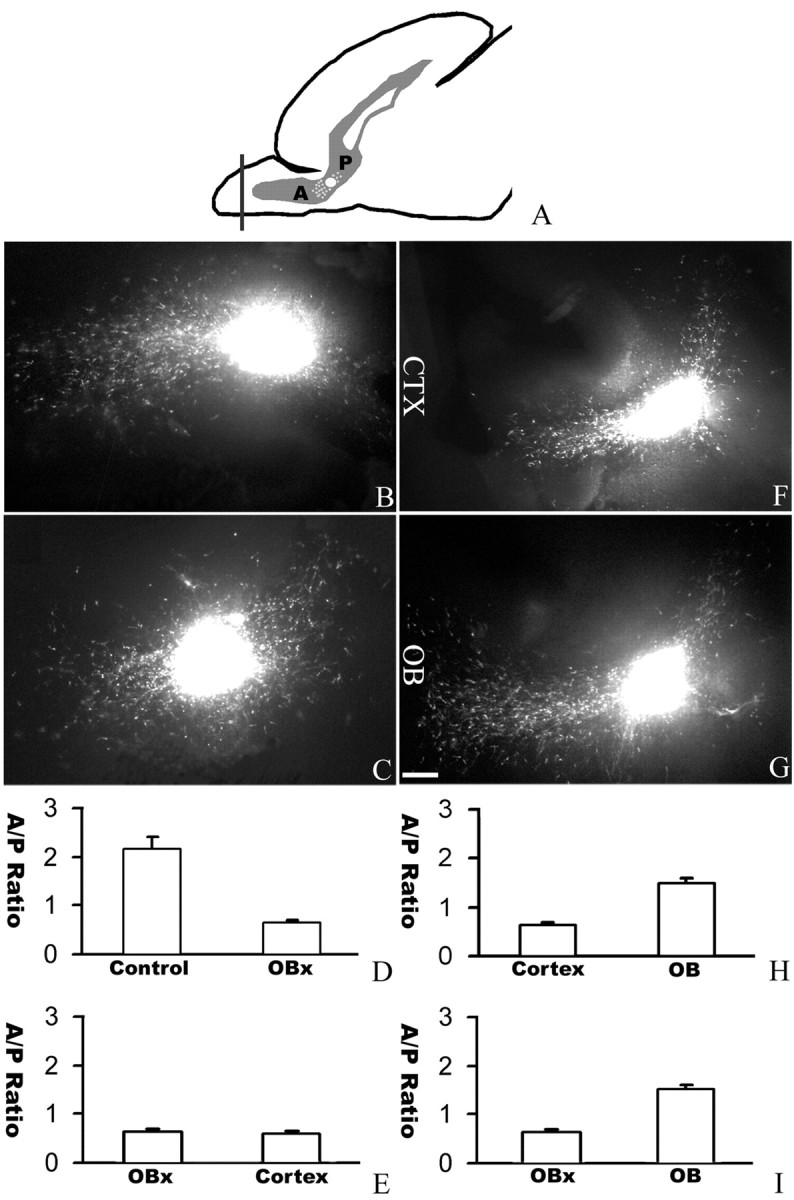
An essential role of the OB in directing neuronal migration in the RMS. A, A diagram of the sagittal section of a neonatal forebrain showing the position of DiI insertion at the juncture of the anterior (A) and posterior (P) parts of the RMS. B, In a normal slice, more cells migrating anteriorly toward the OB. C, When the rostral end of the OB was removed, the distribution of migrating cells was changed with reduced anterior migration and increased posterior migration. D, Effect of OB removal in directing neuronal migration in the RMS. The cell numbers were counted from 20 intact sagittal slices and 32 sagittal slices without the rostral end of the OB. E, Cortical transplants could not functionally replace the rostral end of the OB in directing anterior migration. The cell numbers were counted from 32 slices with the rostral end of their OB removed and nine cortical transplants placed at the rostral end of the OB. F, When a cortical transplant was used to replace the rostral end of the OB, the distribution of migrating cells could not be rescued. G, When an OB transplant was placed at the rostral end of the OB after the original rostral end of the OB was removed, the distribution of migrating cells was changed with more cells migrating anteriorly toward the OB. H, I, The cell numbers were counted from 32 sagittal slices without the rostral end of the OB, nine cortical transplants, and seven OB transplants placed at the rostral end of the OB. CTX, Cortex; A/P, anterior/posterior. Scale bar, 100 μm.
