
Figure 3.
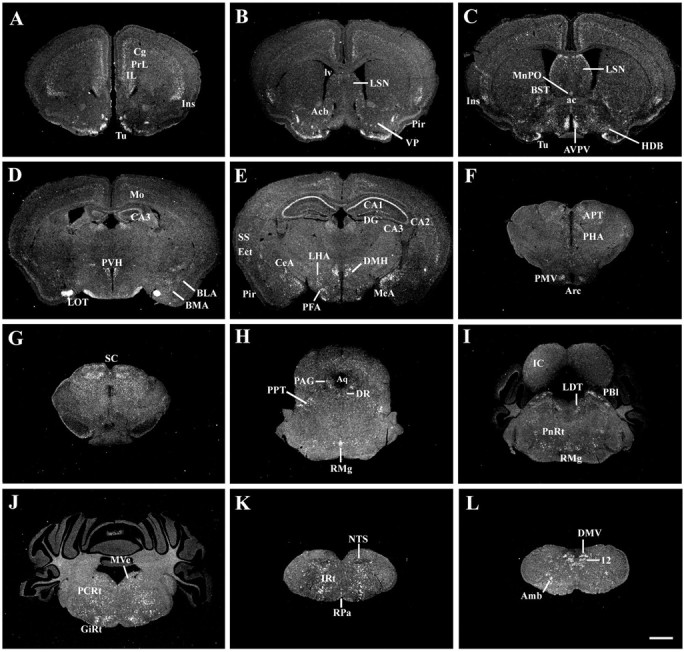
A series of low-power photomicrographs summarizing MC4-R mRNA expression sites across the WT mouse brain. Brain sections are arranged in a rostral-to-caudal manner (A-L). Scale bar (in L), 1 mm (A-L). (For list of abbreviations, see Fig. 2 legend.)