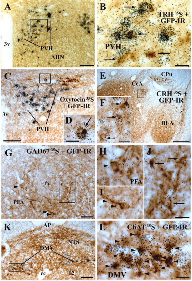
Figure 6.

A series of photomicrographs showing chemical profiles of MC4-R/GFP cells. The GFP-IR cells contain brown cytoplasmic reaction product. The cells containing clusters of grains were hybridized with a 35S-labeled probe for TRH in the PVH (A,B), oxytocin in the PVH (C D), CRH in the central amygdaloid nucleus medial division (CeM) (E,F), GAD67 in the PFA (G-J), or ChAT mRNA (K,L). Boxed areas in A,C,E,G,K are magnified in B, D, F, J, and L, respectively. A cell indicated by an arrowhead and another cell by two arrowheads in G are magnified in H and I, respectively. Arrows and arrowheads indicate double-labeled cells. Scale bars: A,C,K, 100 μm; E, 200 μm; B,D,F, H-J,L, 20 μm; G, 50 μm.
