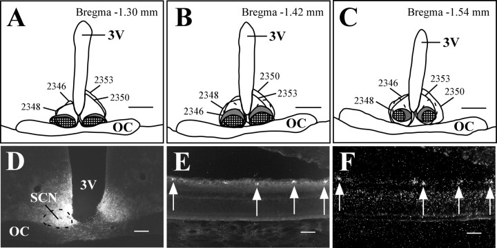
Figure 2.
Melanopsin is expressed in the majority of RGCs that project to the SCN. A-C, Camera lucida drawings of coronal brain sections from FG-injected animals. The ventrolateral SCN is checkered, the dorsomedial SCN is colored gray, and the dashed outline dorsal to the SCN indicates the part of the vSPZ that receives relatively sparse retinal input. Smoothly drawn lines indicate injection sites. All injections were made in the right SCN, but some are transposed to the left side for clarity. D-F, Case 2348. Injections of FG in the SCN (D) resulted in retrogradely labeled RGCs in the contralateral eye (E) that were also positive for melanopsin transcript (F). Arrows indicate double-labeled cells. 3V, Third ventricle; OC, optic chiasm. Scale bars: A-C, 500 μm; D, 200 μm; E, F, 50 μm.

