
Figure 1.
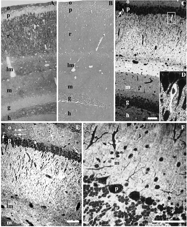
Hippocampal (CA1 and dentate gyrus) semithin cryosections labeled for PrPC with Fab D18. Labeling is concentrated in the stratum oriens (o), stratum radiatum (r), and lacunosum-moleculare (lm); less labeling is seen in the stratum moleculare (m) and hilus (h) of the dentate gyrus. Cell bodies in pyramidal (p) and granule (g) layers are free of labeling, with the exception of rare cells (D). A, Hippocampus of a wt FVB mouse. Fab D18 was used with a secondary polyclonal antibody and protein A-gold (5 nm) that was subsequently visualized by silver enhancement (Aurion). B, Hippocampus of a Prnp0/0 mouse. Labeling is the same as in A. No positive signal is visible. Sections are counterstained with Giemsa. C, Hippocampus of a wt FVB mouse. Fluorescent labeling shows a labeling pattern similar to A. The arrow points to a cell with intense PrPC labeling. The box shows the cell enlarged in D. D, A magnified view of a cell with high PrPC content. E, Hippocampus (CA1 area) of a 4053 mouse that overexpresses PrP, showing a pattern of PrPC labeling similar to A and C. Arrows point to the cells with an abundance of PrP in the cytoplasm. F, Cerebellum of a wt FVB mouse. No cells labeling positively for PrPC in the cytoplasm were found in the cerebellum. Purkinje cells (p) are free of intensive cytosolic labeling. Only faint punctate labeling is visible. Scale bars, 100 μm.