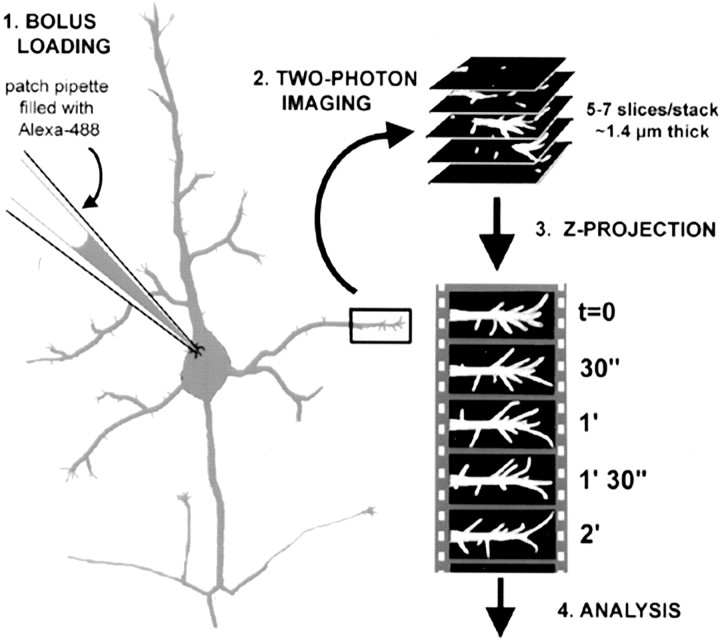
Figure 1.
Bolus of neurons with Alexa-488 and two-photon imaging of filopodia. Step 1 (bolus loading): cortical pyramidal neurons in layer 5 were identified in acute slices from early postnatal mice under differential interference contrast optics and then patched with pipettes containing 2 mm Alexa-488. This dye diffuses quickly (<5 min) throughout the entire cell. Step 2 (two-photon imaging): selected dendrites were imaged with a custom made two-photon microscope using 800 nm excitation light. Twenty stacks, each composed of 5-7 confocal slices (1-1.4 μm apart) in the XY plane, were acquired every 30 sec. Step 3 (Z-projection): using ImageJ software, the individual slices for each time point were projected along the z-axis into a single image. Ten-minute-long (20 time points) time-lapse movies of dendritic protrusions were, thus, generated. Step 4 (analysis): the lengths and density of dendritic protrusions were measured using the ROI tool in ImageJ (see Materials and Methods and Fig. 3).

