
Figure 10.
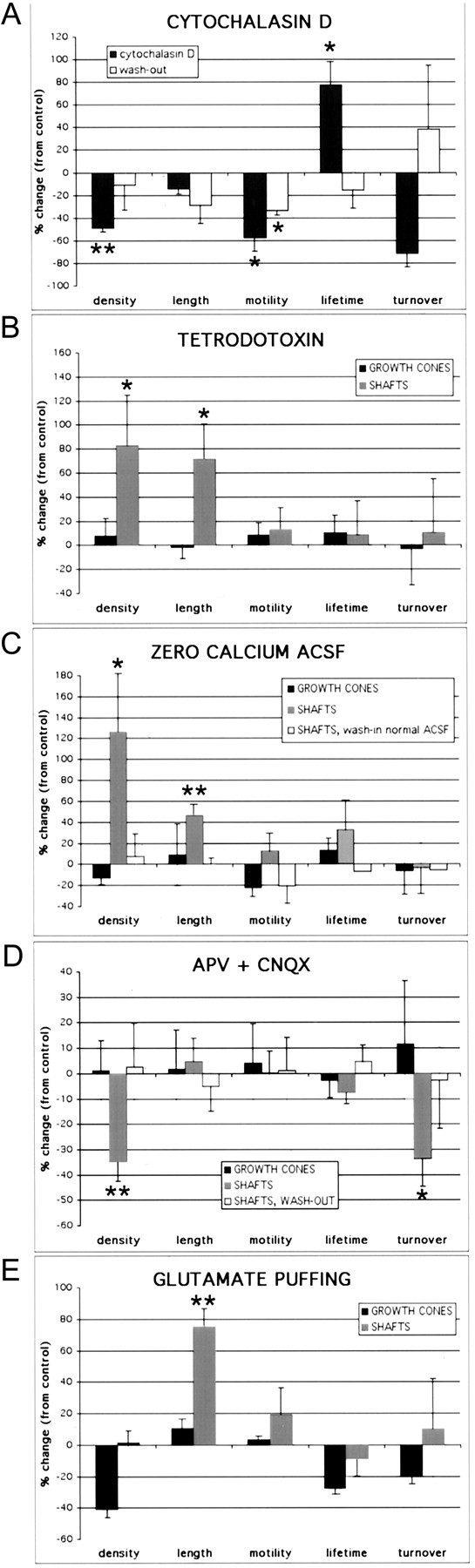
Dendritic shaft filopodia, but not growth cone filopodia, are regulated by neronal activity. A, Bath application of 1 μg/ml cytochalasin D, an inhibitor of actin polymerization (n = 46; four experiments), resulted in an ∼50% decrease in density (p = 0.004), an ∼55% decrease in motility (p = 0.011), and an ∼75% increase in lifetime (p = 0.016) of dendritic protrusions. Shaft and growth cone protrusions were equally affected. An ∼15% decrease in the length of filopodia approached, but did not reach significance (p = 0.074), The effect on density and lifetime was reversible after washing out the drug, because there were no significant differences between control and washout conditions (p > 0.26; n = 90; three experiments). This wash-out effect required several hours (the data come from dendrites imaged on average ≥4 hr after washout of cytochalasin D). B, Blocking neuronal activity with 1 μm TTX (n = 219; five experiments) resulted in an ∼85% increase in the average density of shaft filopodia (p = 0.04) and an ∼75% increase in the average length of dendritic shaft filopodia (p = 0.05). Neither the density nor the length of dendritic growth cone filopodia were significantly affected by TTX (p = 0.70 and p = 0.69, respectively). Motility and lifetime of filopodia were unaffected by TTX in either dendritic shafts (p > 0.21) or growth cones (p > 0.54). C, Blocking neuronal activity with calcium-free ACSF (n = 350; six experiments) also resulted in an ∼125% increase in the average density (p = 0.05) and an ∼45% increase in the average length (p = 0.004) of filopodia in dendritic shafts. Just as with TTX, neither the density nor the length of dendritic growth cone filopodia were significantly affected in the presence of zero-calcium ACSF (p = 0.52 and p = 0.88, respectively). Motility and lifetime of filopodia were similarly unaffected by zero-calcium ACSF in either dendritic shafts (p > 0.23) or growth cones (p > 0.32). The effects of zero-calcium ACSF on dendritic shaft filopodia density and length were reversible ≥30 min after washing in normal ACSF containing 2 mm CaCl2 (p > 0.28 compared with control; N = 3 successful washouts; n = 59). D, Blocking ionotropic glutamate receptors for 15-30 min with 20 μm CNQX and 40 μm d-APV resulted in an ∼35% decrease in both the density (p = 0.006) and the turnover (p = 0.04) of shaft filopodia (N = 8; n = 319) but had no effect on length, motility, or lifetime of shaft protrusions (p>0.15). CNQX/APV had no effect on growth cone filopodia (p>0.56; N = 5; n = 164). These effects of CNQX/APV on dendritic shaft filopodia were reversed 30-60 min after washing out the drug (p > 0.63 compared with control; N = 4 successful washouts; n = 116).E, PicoSpritzer application of 100-200 μm glutamate resulted in an ∼75% increase in the length of shaft filopodia (p = 0.004; N = 5; n = 251) but did not significantly affect the other parameters (p > 0.25). A trend toward lower density of growth cone filopodia was also observed (p = 0.055; N = 2; n = 76). The histograms in A-E reveal percentage differences compared with control dendrites imaged before the application of the drugs or zero-calcium ACSF. Error bars represent the SEM, using the number of experiments (N), rather than the number of protrusions. *p < 0.05; **p < 0.01.