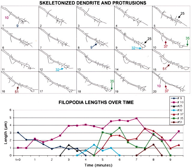
Figure 3.
Dendritic filopodia are highly motile protrusions in early postnatal development. Top, Skeleton representation of all 20 frames of the movie shown in Figure 2. Individual protrusions were traced in ImageJ (see Materials and Methods) in all 20 frames of each time-lapse movie. Skeleton versions of movies of filopodia and other protrusions were drawn for all dendrites imaged in this study throughout development from P2 through P12 (a total of 56 dendrite segments and 1008 filopodia), to measure their lengths using custom-written macros in ImageJ. A few representative filopodia are labeled; arrows point to the first and last frame in which those filopodia can be distinguished. Bottom, Graph displaying the lengths of the representative filopodia mentioned above over the same 10-min movie. Most filopodia appear and disappear over the 10-min imaging period (e.g., filopodia #25, #32, and #51). At these early ages, only rare filopodia were present throughout the length of the movie (e.g., filopodium #10).

