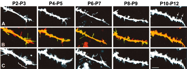
Figure 5.
Dendritic filopodia are developmentally regulated; dendritic growth cones disappear after P5. The first row (A) shows representative dendritic segments of cortical neurons of mice at P2 through P10 (see movies 3-7 in supplementary data, available at www.jneurosci.org). The second row (B) shows composite views of the first (red) and last (green) frames of the 10-min time-lapse movies of the dendrites. The third row (C) shows a collapsed view of all 20 frames of the 10-min movie at each postnatal age. Scale bar, 5 μm. These panels show four results. First, filopodia are longer and more densely packed in dendritic growth cones than in shafts. Note that dendritic growth cones (bracket in C) are only apparent at P2-P3 and P4-P5. Second, dendritic protrusions become progressively more densely packed throughout early postnatal development and become shorter in dendrite tips. Third, as judged by the overlap between first and last time frames in each movie (second row), a switch from highly motile, transient filopodia to more stable, immobile protrusions occurs throughout early postnatal development. Fourth, spine-like protrusions, with bulbous swellings at their tips (heads), begin to appear at P10-P12 (arrows in A).

