
Figure 6.
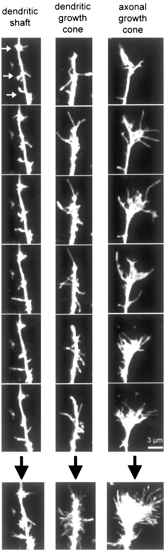
Differences between filopodia in axonal and dendritic growth cones and between filopodia in dendritic growth cones and in dendritic shafts. Left, Example of a typical dendritic shaft at P2-P5. Note that filopodia are shorter than in both axonal growth cones and dendritic growth cones, and they are less densely packed. There are also three hotspots of filopodia activity (white arrows). These hotspots were frequently seen in young dendrites but almost never after P6. Middle, Example of a typical dendritic growth cone at P2-P5. Note how individual filopodia are easily identified (no webbing) and that filopodia aim in all directions, including away from the tip of the dendrite. Right, Example of a typical axonal growth cone at P2-P5. Note the long filopodia and the webbing between filopodia. Almost all filopodia are oriented at acute angles toward the tip of the axon. The different rows show different time points in the 10-min time-lapse movies. The last row (below the black arrows) shows collapsed sums of all 20 individual time points from each 10-min movie. Scale bar, 3 μm.