
Figure 8.
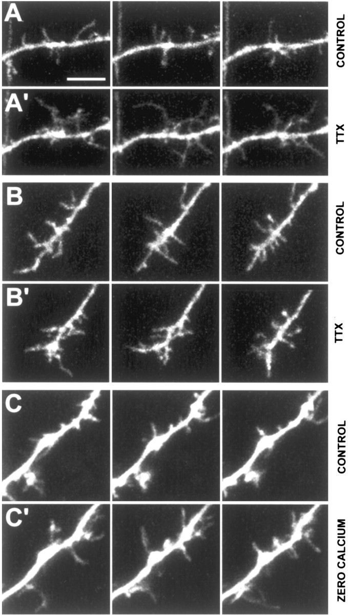
Blockade of global synaptic activity results in longer, more motile filopodia. A, Dendritic shaft from a control neuron at P3 in control ACSF (2 mm CaCl2). The left, middle, and right panels in each row show representative snapshots of dendritic filopodia in the beginning, middle, and end of the 10-min movies. Scale bar, 5 μm (for all panels). A′, Same dendritic shaft segment ∼7 min after continuous bath application of 1 μm TTX. Note the dramatic elongation of many filopodia in three different frames of a 10-min time-lapse movie. B, Dendritic growth cone from a control neuron at P4 in control ACSF (2 mm CaCl2). B′, Same dendritic growth cone ∼30 min after continuous bath application of TTX. Note that there is no change in the density or length of growth cone filopodia. C, Dendritic growth cone from a control neuron at P3 in control ACSF (2 mm CaCl2). See movie 8 in supplementary data, available at www.jneurosci.org. C′, Same dendritic growth cone ∼25 min after washing in zero-calcium ACSF. Note the dramatic elongation of many filopodia in three different frames of a 10-min time-lapse movie. This effect was reversible because filopodia lengths and motility recovered after washout. See movie 9 in supplementary data, available at www.jneurosci.org.