
Figure 9.
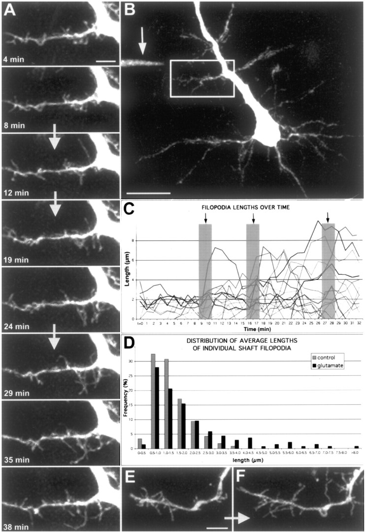
Focal application of glutamate results in rapid elongation of a subset of filopodia. A, Time-lapse movie of apical dendrite from a layer 5 pyramidal neuron at P4 (see movie 10 in supplementary data, available at www.jneurosci.org). Images were acquired every 60 sec, and representative time points are shown. The white arrows separating certain frames represent the times when three puffs (200 msec, ∼20 psi) of 100 μm glutamate were delivered, corresponding to frames 10, 17, and 28. Note that by the end of the movie, many filopodia have grown to lengths >5 μm. Scale bar, 5 μm. B, In this low magnification view of a pyramidal neuron, the white box delineates the region in which the movie in A was obtained. The arrow points to the location of the pipette tip containing 100 μm glutamate (and Alexa-488). Scale bar, 20 μm. C, Graph displaying the lengths of 45 representative filopodia from the 40-min movie shown in A. Black arrows and gray columns designate the three times when glutamate was puffed (200 msec puffs). Note that during the first 10 frames of the movie filopodia lengths never surpass 4 μm, whereas after just two puffs of glutamate, a subset of filopodia have reached lengths well above 4 μm. The effect of glutamate was quite rapid as shown by the sudden increases (within 1 min) of some filopodia after individual puffs. D, Frequency distribution of filopodia lengths before and after glutamate application from five separate experiments. The gray histograms represent pooled data from 117 filopodia analyzed in the first 10 frames of each movie, whereas the black histograms represent data from 136 filopodia analyzed during 10 consecutive frames after at least two puffs of glutamate. Note that glutamate induced growth to lengths >4 μm in a minority (∼10%) of filopodia, rather than a small increase in the length of all the filopodia. E, F, Another example of the effects of glutamate puffing on a P2 dendrite. The pipette was located ∼50 μm away, up and to the left of the dendrite growth cone. E is a representative frame of the dendrite with a growth cone in normal conditions. F is the same dendrite ∼30 min after three puffs of 200 μm glutamate. Scale bar, 5 μm.