
Figure 8.
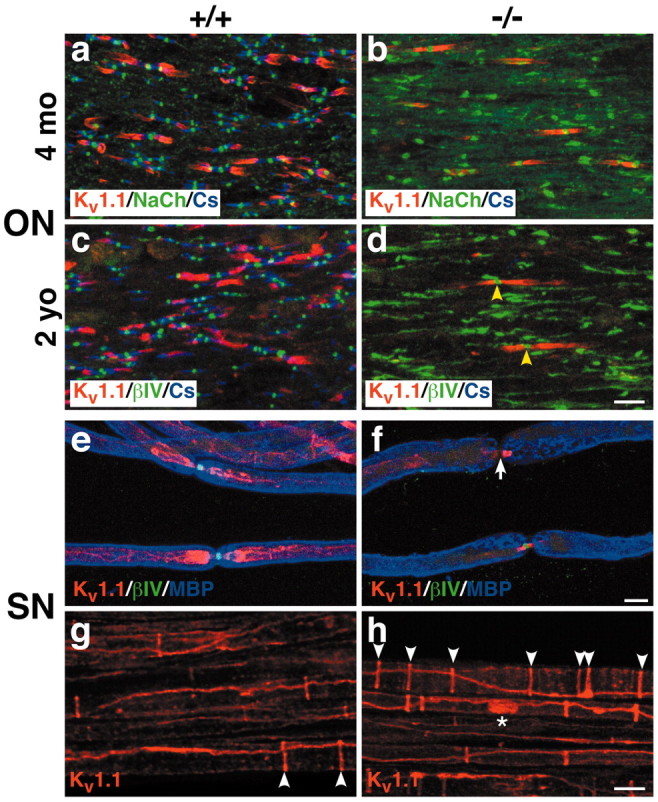
Potassium channel distribution is aberrant in Caspr-deficient mice. Optic nerve sections of 4-month-old (a,b) and 2-year-old (c,d) optic nerves from wild-type (+/+) and Caspr mutants (-/-) were triple stained for Kv1.1 (red), Caspr (blue), andβIV spectrin (green). Kv1.1 is present in many of the juxtaparanodes of the wild type (a,c) but only a few of the paranodes of the Caspr mutants (b,d), particularly at 2 years. Kv1.1 expressed in the paranodes of the mutants frequently flanks well delineated nodes; two examples are indicated (d; yellow arrowheads). Sciatic nerves from wild-type (e,g) and Caspr mutant (f,h) littermates were triple stained for Kv1.1 (red),βIV spectrin (green), and MBP (blue) (e,f) or Kv1.1 (red) alone (g,h). Kv1.1 is expressed in the juxtaparanodes of wild-type mice (e) and the paranodes of Caspr mutants (f). Deficient Kv 1.1 expression in the paranodes of Caspr mutants is associated with attenuatedβIV spectrin at the node (f, white arrow). There are more Schmidt-Lanterman clefts in the Caspr mutant (h, white arrowheads) than in the wild type (g, white arrowheads). Kv1.1 is also occasionally expressed in a band along the axon (h, asterisk). Scale bars, 10 μm.