
Figure 2.
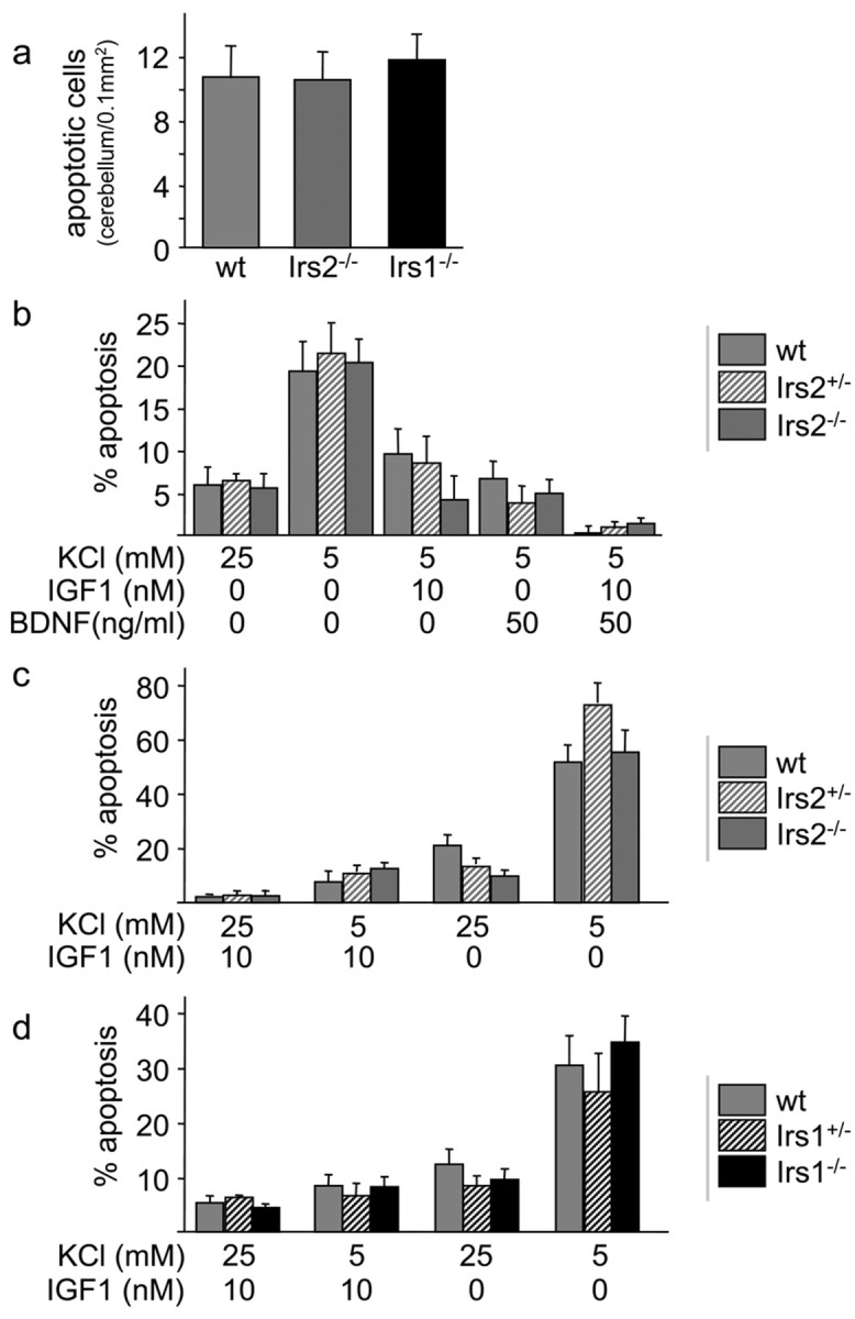
Apoptosis in brains and cultured neurons from mice lacking IRS proteins. a, TUNEL assay of the cerebellum at P6 reveals no differences between Irs2-/-, Irs1-/-, and wild-type (wt) mice. Quantification of apoptotic nuclei showed the same number per area in all genotypes. b, IGF1 and BDNF protect against neuronal apoptosis in the absence of Irs2. Cerebellar granule cells (P5) were cultured for 4 d in medium containing normal serum. Neurons were then incubated for an additional 24 hr in serum-free media containing 5 mm KCl, 5 mm KCl plus 10 nm IGF1, 5 mm KCl plus 50 ng/ml BDNF, or 25 mm KCL. Cells were stained with Hoechst 33342 and counted. Percentage apoptosis was calculated (n = 3 per genotype and experiment; 3 independent experiments showed similar results). c, Cerebellar granule cells from Irs2+/- × Irs2+/- litters (Irs2+/- or Irs2-/- cells) were cultured for 4 d, followed by incubation for 24 hr in serum-free media containing 25 mm KCl plus 10 nm IGF1 (+KCl, +IGF1), 10 nm IGF1 (-KCl, +IGF1), 25 mm KCl (+KCl, -IGF1), or minimal medium (-KCl, -IGF1) (n = 2 per genotype and experiment; 3 independent experiments showed similar results). d, Cerebellar granule cells from Irs1+/- × Irs1+/- litters (Irs1+/- or Irs1-/- cells) were cultured for 4 d, followed by incubation for 24 hr as described in c.