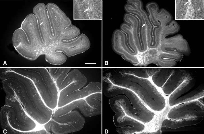
Figure 1.
Mouse developmental myelination in vivo. Parasagittal slices of cerebella at P3 (A), P7 (B), P10 (C), and P21 (D) immunostained with an antibody against MBP. A, At P3, numerous MBP-positive cells are present in the area of the deep nuclear neurons and few in the white matter of the folia (see high magnification in the inset). B, At P7, myelinated segments can be observed in the white matter of the folia (see high magnification in the inset). C, At P10, almost all the axes of the folia are myelinated, and some myelin segments start to appear in the granule cell layer. D, At P21, the cerebellum is almost fully myelinated. Note that MBP-positive segments are present at high density in the granular cell layer. The arrows in A and B indicate to the areas presented at higher magnification in their respective insets. Scale bar: A-D, 220 μm; insets, 75 μm.

