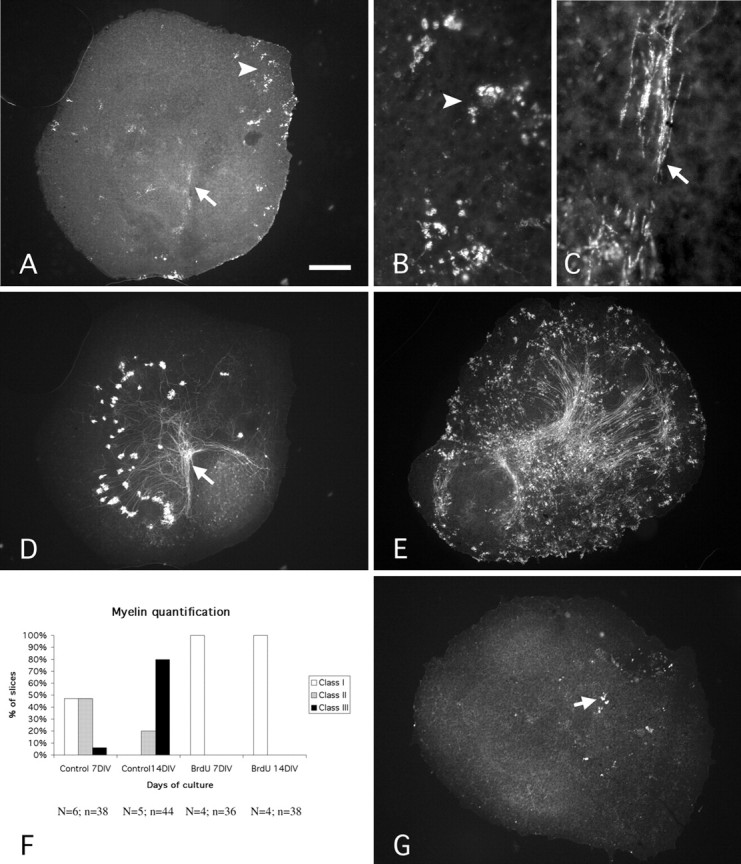
Figure 2.

Myelination and inhibition of myelination in vitro. A-D, Photomicrographs of a P0 cerebellar slice maintained 7 DIV and double labeled with antibodies against MBP (A-C) and CaBP (D). MBP-positive oligodendrocytes (arrowhead) and segments (arrow) are present (A). Note that the packs of MBP-positive segments are numerous in the area of deep nuclear neurons revealed by the presence of numerous Purkinje cell axon terminals (A, D, arrows). B, Higher magnification of an area of A showing MBP-positive oligodendrocytes. Arrowheads in A and B point to the same area. C, Higher magnification of the area indicated by the arrow in A, showing MBP-positive segment packs. Arrows in A and C point to the same area. E, Photomicrograph of a P0-14 DIV slice immunostained with an antibody against MBP showing the high number of MBP-positive cells and segments in comparison with the P0-7 DIV slice (A). F, Quantitative evaluation of myelination. The three classes of slices (I, II, and III) were defined according to the number of MBP-positive cells and segments. Class I included the slices with a small number of oligodendrocytes and of packs of internodes (<15 of each; G). Class II included slices containing >16 oligodendrocytes or 16 internode packs but <20 packs of internodes (A). Class III included slices containing >21 packs of internodes (E). N, Number of animals; n, number of slices. G, Photomicrograph of a P0-14 DIV BrdU-treated slice immunostained with an antibody against MBP. Note that only a few oligodendrocytes (arrow) can be observed. Scale bar: A, D, E, G, 270 μm; B, C, 30 μm.
