Figure 7.

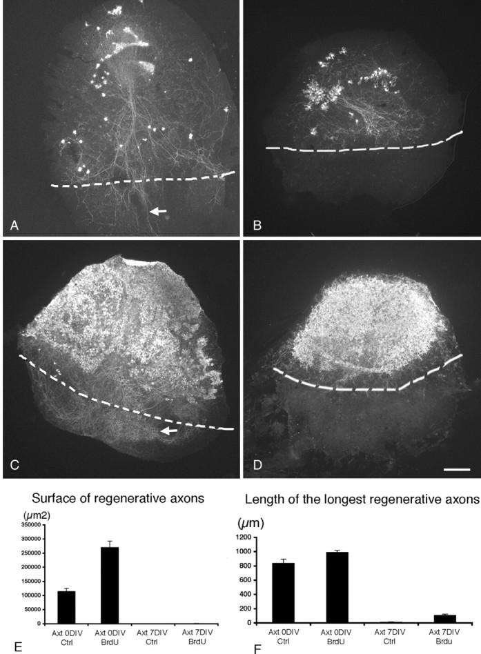
The absence of myelin does not favor axonal regeneration of Purkinje cells. Photomicrographs showing cocultures of the dorsal region of wild-type P0 mouse cerebellar slices with the ventral region of P0 cerebellar slices taken from a CaBP null mutant immunostained with CaBP antibodies. A, B, Control slices. C, D, BrdU-treated slices. The limits between the two cultures are indicated by dashed lines. A, The axotomy and the coculture were performed at 0 DIV. Note that the surviving Purkinje cells regenerate their axons after 7 DIV (A, arrow). B, The axotomy and the coculture were performed at 7 DIV; the few surviving Purkinje cells did not regenerate their axons after 14 DIV. C, D, BrdU-treated cocultures. Note that BrdU treatment greatly improves the survival of axotomized Purkinje cells at 0 DIV (C) and at 7 DIV (D) and that, like control cocultures, Purkinje cells are able to regenerate their axons when axotomy is performed at 0 DIV (C, arrow) but not when axotomy is performed at 7 DIV (D). E, F, Quantitative analysis of Purkinje cell regeneration (axon length and surface covered by regenerative axons). On each section, the surface covered by the regenerative axons (under the limit of the coculture; A-D, dashed line) and the longest regenerative axon were measured. E, Mean surface covered by regenerative axons. F, Mean length of the longest regenerative axon per slice in BrdU-treated slices and in control slices (Ctrl) axotomized (Axt) at 0 DIV or at 7 DIV. The measure of the surface covered by regenerative axons takes into account both regenerative axon lengths and numbers (E). Scale bar, 220 μm.
