
Figure 5.
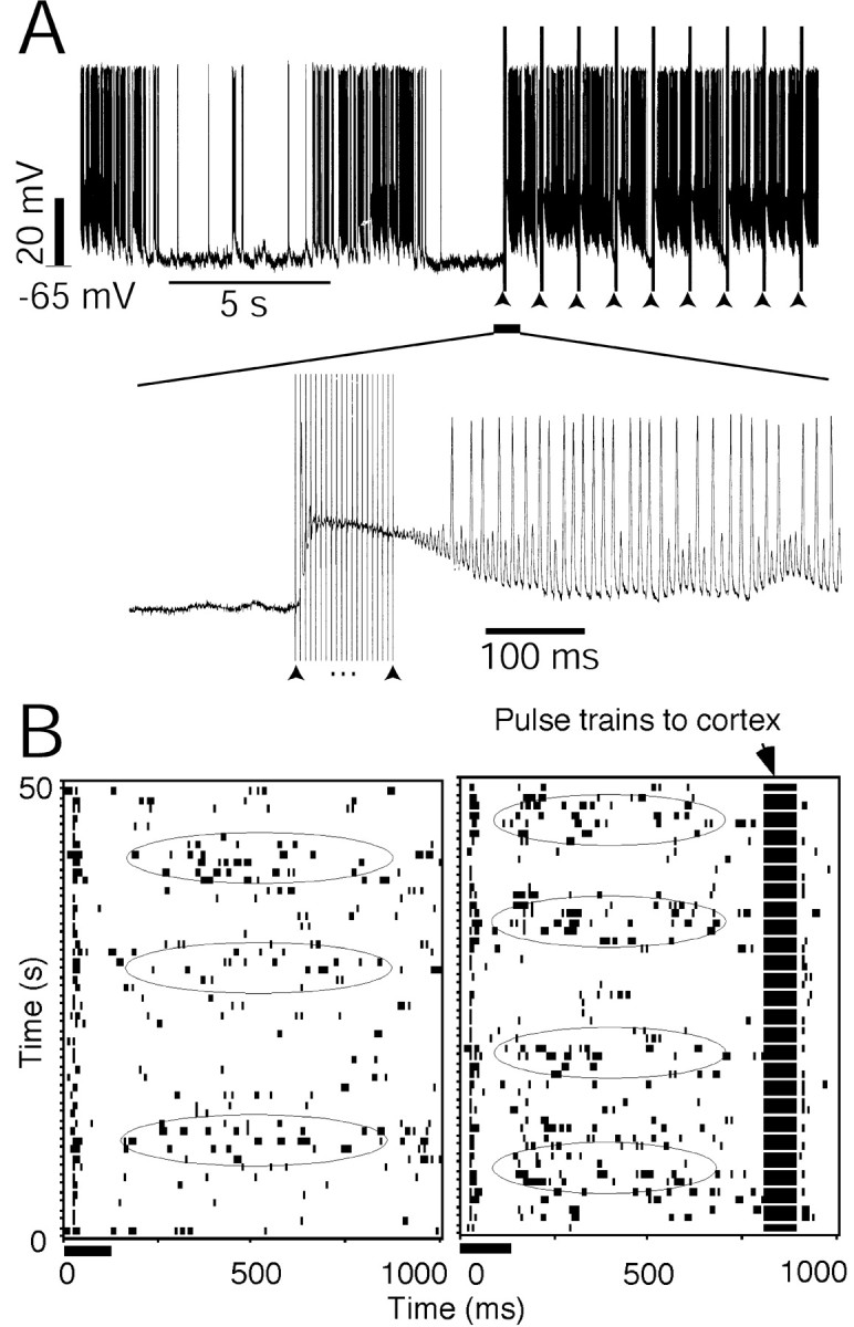
Thalamic oscillation altered by the corticofugal modulation. A, An intracellular recording shows that the thalamic oscillation was terminated, whereas the electrical stimulation of the auditory cortex was turned on. The bottom panel shows the time-axis expanded from when electrical pulses were applied to stimulate the cortex. Arrowheads indicate the pulse trains of the electrical currents applied to the auditory cortex. The electrical stimulation current was 200 μA. B, Raster displays show the neuronal responses to a repeated auditory stimulus. The neuron was located in the caudomedial nucleus of the non-lemniscal MGB. Left, Only auditory stimulus was given to the subject; right, the auditory cortex was activated 100 msec before the auditory stimulus was delivered to the subject. The rhythm became faster when the cortex was stimulated. The ellipses indicate the peak phases of the slow oscillation waves. The electrical stimulation current was 100 μA.