
Figure 1.
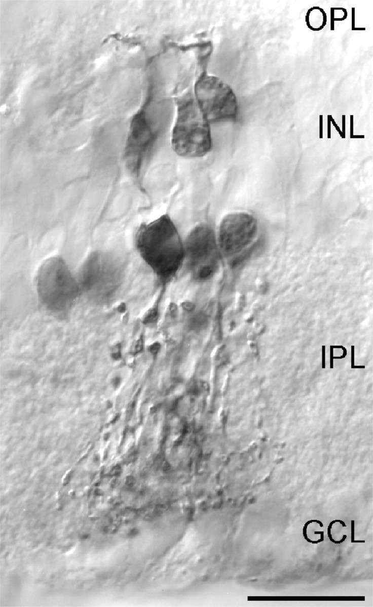
Micrograph of a retinal slice showing neurobiotin staining of a group of coupled AII (inner IPL) and ON-CB cells (outer IPL) after filling a single AII amacrine cell via the patch pipette. The strongly stained soma located in the innermost part of the INL is the patched cell. Robust dendrites that constitute the lobular appendages of AII cells are observed in sublamina a of the IPL, whereas the more bushy arboreal dendrites are observed in sublamina b. Scale bar, 10 μm.