
Figure 2.
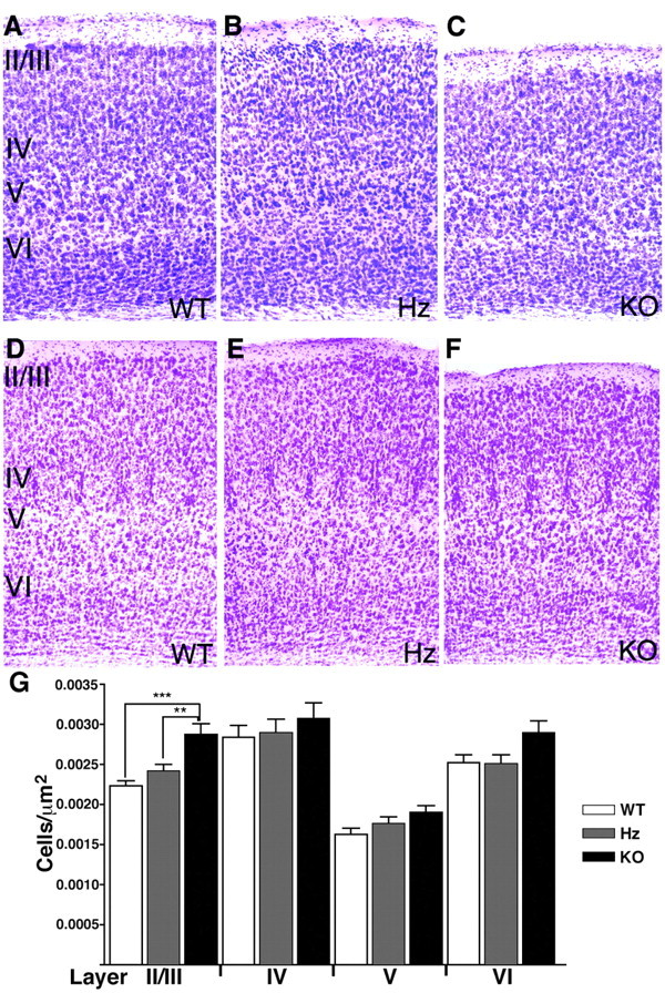
Cortical thinning in Emx-BDNFKO mice. Cresyl violet-stained 50 μm coronal sections of visual (A-C) and somatosensory (D-F) cortices from 5-week-old WT, Hz, and KO mice. Cortical thickness is reduced in KO mice, but the cytoarchitecture appears generally normal and barrels are apparent in somatosensory cortex (D-F). G, Neuronal density in visual cortex of 5-week-old mice (n = 3 mice per genotype). Neurons are distributed more densely in layer II/III of KO mice (p < 0.05).