
Figure 3.
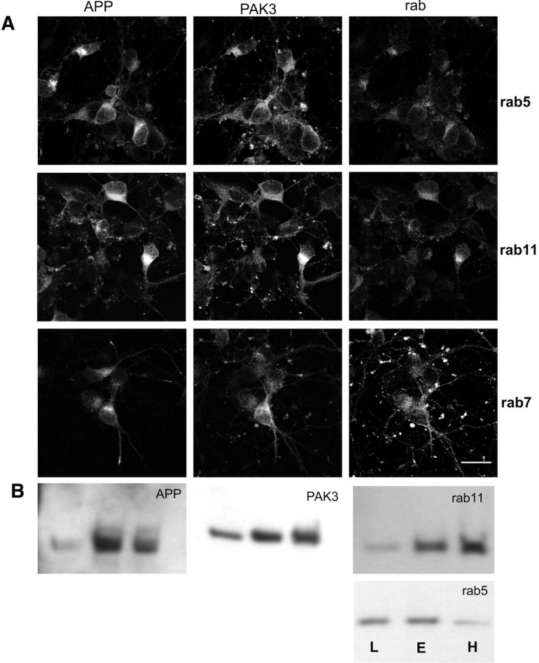
APP and PAK3 colocalize in rab5- and rab11-positive structures in neurons. A, Neuronal cultures were infected with both HSV-PAK3 and HSV-APP-695. The first column of cells represents APP immunofluorescence, the middle column PAK3 immunofluorescence, and the last column rab5, rab7, or rab11 immunofluorescence, as indicated by the row label. Note overall colocalization of APP and PAK3. Rab11 shows slightly more specific colocalization with APP than does rab5. Rab7 localization shows minimal overlap with that of APP or PAK3. Scale bar, 20 μm. B, Immunoblots of endosomal fractionation of rat brain lysates demonstrates that PAK3 and APP cofractionate into rab-11 and rab-5 positive fractions. The three lanes of each blot are designated L, E, and H: L, the membrane fraction that is enriched in late endosomes; E, the membrane fraction that is enriched in early endosomes; H, the fraction that contains heavy membranes.