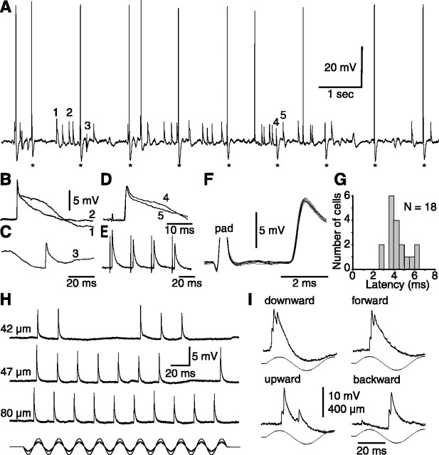
Figure 2.
Unitary character of whisker-evoked EPSPs in barreloid cells. A, Trace shows a short episode of spontaneously occurring and electrically evoked EPSPs (asterisks, follicular stimulation) in a D2-responsive relay cell. Events labeled 1-4 in A are magnified in B-D. Superimposed traces (B, D) show that EPSPs have similar rise times but variable falling phases. Note that the late component is much reduced when EPSPs are superimposed onto an IPSP (C). Time scale in C also applies to B. The thick trace in D is the stimulus-evoked response. E, Trace shows the depression of lemniscal EPSPs evoked in a C3-responsive cell by repetitive electrical stimulation of the whisker follicle. F, Superimposed traces (n = 10) of EPSPs evoked in a C2-responsive cell by suprathreshold electrical stimulation of the follicle (pad). Note the very small jitters of the bisynaptic responses. G, Shown are the shortest latencies of EPSPs evoked in 18 VPM cells by electrical stimulation of the whisker follicles. H, All-or-none occurrence of EPSPs evoked in a C2-responsive cell by straddling threshold (42 and 47 μm) and suprathreshold deflections (80 μm). I, Bursts of unitary EPSPs evoked at stimulus onset by deflecting whisker B4 in different directions. All of the cells but the one in I were classified as single-whisker neurons.

