
Figure 4.
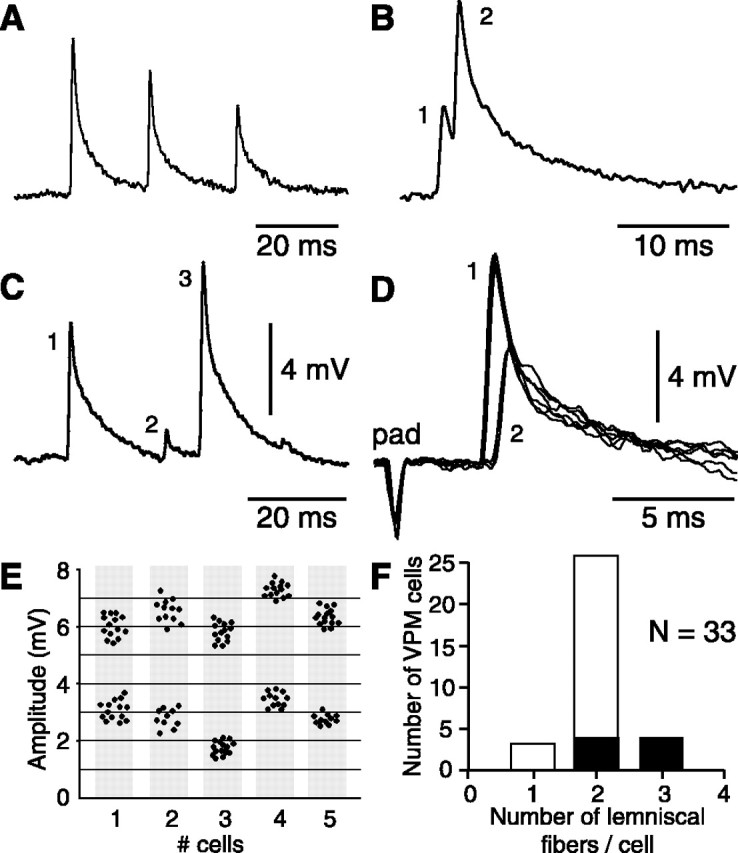
Criteria used to estimate the number of lemniscal fibers that contact single barreloid cells. A, Trace shows the synaptic depression observed when whisker-evoked EPSPs occurred at short intervals. In this case, it was inferred that the EPSP sequence resulted from the firing of a single presynaptic fiber. B, C, Traces show cases that break the rule of synaptic depression. To generate the sequences shown in B and C, two and three presynaptic fibers are required, respectively. D, Additional evidence for the convergence of lemniscal fibers on the same barreloid cells was obtained after electrical stimulation of the follicle (pad) or of the medial lemniscus. Superimposed traces (n = 6) show single-fiber EPSPs of different amplitudes evoked at slightly different stimulus intensities. E, Scatter plots summarize the amplitude distribution of evoked EPSPs in five additional cells (each plot contains 20-30 measures). F, On the basis of these criteria, the histogram shows the number of lemniscal fibers that contact each of the 33 barreloid cells analyzed. Filled bars represent cells that were strongly driven by multiple whiskers, and open bars represent single-whisker units.