Figure 5.

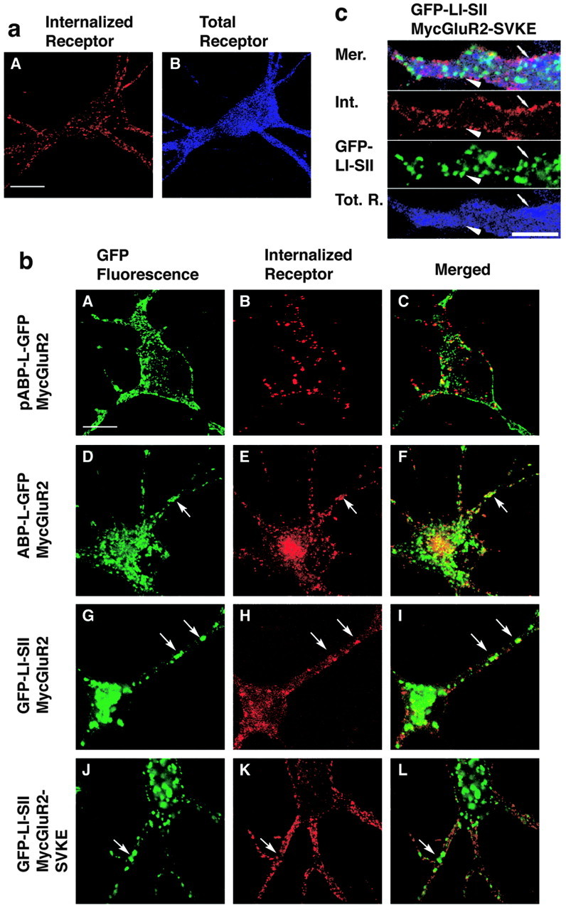
Internalized MycGluR2 colocalizes with pABP-L-GFP, ABP-L-GFP, and GFP-LI-SII. a, Neurons were infected with Sindbis viruses expressing MycGluR2 alone. After 24 hr of infection, living cells were incubated with monoclonal anti-Myc antibody, followed by acid stripping and immunostaining. The total receptor was probed by the polyclonal anti-Myc antibody. Scale bar, 20 μm. b, Neurons coexpressed MycGluR2 with ABP or ABP fragments as indicated. All of the ABPs were seen by GFP fluorescence (A, D, G, J). MycGluR2 and its mutant were detected as red (internalized; B, E, H, K) or blue (total proteins; C, F, I, L). MycGluR2 (B) was detected on the plasma membrane and in spines (A), which does not colocalize exactly with pABP-L-GFP, when coexpressed with pABP-L-GFP. However, it coclustered with ABP-L-GFP (compare D, E) or with GFP-LI-SII (compare G, H), when coexpressed with ABP-L-GFP or GFP-LI-SII, respectively. As a control, Myc GluR2-SVKE was also coexpressed with GFP-LI-SII (J-L). Internalized MycGluR2-SVKE did not cocluster with GFP-LI-SII (compare J, K). Arrows indicate the sites at which MycGluR2 and ABPs colocalized or not. c, Dendritic expression of GFP-LI-SII and MycGluR2-SVKE at higher magnification. Panels are merged (Mer.) images, internalized MycGluR2-SVKE (Int.), GFP-LI-SII, and total receptor (Tot. R.). Note that the GFP-LI-SII clusters (arrowhead) and internalized receptor (small arrow) do not colocalize with each other. Scale bar, 10 μm.
