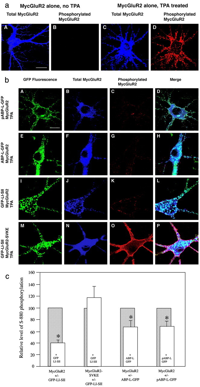
Figure 6.

Ser880 phosphorylation of GluR2 is partially inhibited by coexpression of ABP. a, Hippocampal neurons were infected with Sindbis virus expressing MycGluR2 alone. The effect of TPA was assessed by comparing neurons not treated (A, B) or treated (C, D) with TPA. Total MycGluR2 was detected by antibody against Myc epitope, which is shown in blue (A, C), and the phosphorylated MycGluR2 was probed with antibody against the S880-PO4 phosphorylated peptide and is shown in red (B, D). In the absence of TPA, no signal is detected using antibody against the phosphorylated peptide (B). In the presence of TPA, intensive staining was shown using the same antibody (D). Scale bar, 20 μm. b, Hippocampal neurons were coinfected with Sindbis virus expressing pABP-L-GFP (Figure legend continued.) and MycGluR2 (A-D); ABP-L-GFP and MycGluR2 (E-H); GFP-LI-SII and MycGluR2 (I-L); or GFP-LI-SII and MycGluR2-SVKE (M-P). After 24 hr of infection, neurons were treated with TPA to induce PKC. GFP-tagged ABPs were visualized by fluorescence (A, E, I, M). Total (B, F, J, N) and phosphorylated (C, G, K, O) receptors were detected as shown in a. D, H, L, P, Merged images. Coexpression with MycGluR2 of pABP-L-GFP (C), ABP-L-GFP (G), or GFP-LI-SII (K) reduced phosphorylation relative to MycGluR2 expressed on its own (D). GFP-LI-SII coclusters with the total MycGluR2, which is primarily the unphosphorylated MycGluR2 (compare I, J). In contrast, the level of phosphorylated MycGluR2-SVKE in cells coinfected with MycGluR2-SVKE and GFP-LI-SII is higher (compare O with C, G, K). Scale bar, 20 μm. c, Neurons were infected and treated with TPA as shown in b. The coinfections were with viruses expressing MycGluR2 and GFP-LI-SII; MycGluR2-SVKE and GFP-LI-SII; MycGluR2 and ABP-L-GFP; or MycGluR2 and pABP-L-GFP. Cells were scanned with a confocal microscope, images were quantitated, and the ratio of phosphorylated MycGluR2 (red channel) to total MycGluR2 (blue channel) was determined. The phosphorylated MycGluR2 signals (open bars) were normalized to that obtained from cells with single infection with MycGluR2, scanned from the same coverslip (shaded bars). For each group, 10 or more cells were scanned. Whereas the phosphorylation of MycGluR2 was highly significantly reduced in presence of GFP-LI-SII compared with the control (p < 0.0002), the phosphorylation of MycGluR2-SVKE was not significantly changed in the presence of GFP-LI-SII (p > 0.4). The phosphorylation of MycGluR2 was significantly reduced in the presence of ABP-L-GFP or pABP-L-GFP (p < 0.05). Error bar indicates SEM. *p < 0.05.
