Figure 1.

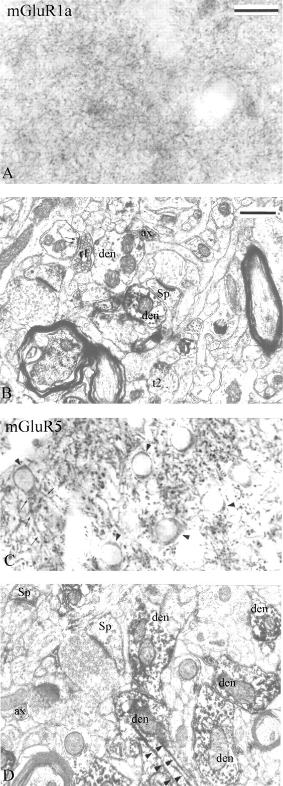
Immunoperoxidase labeling of group I mGluRs in the monkey striatum. A, B, Light and electron micrographs of mGluR1a immunostaining are shown. A, A dense mGluR1a immunoreactive neuropil composed of small punctate elements is depicted. B, Dark DAB reaction product is observed in small dendrites (den), dendritic spines (Sp), unmyelinated axons (ax), and axon terminals (t). Note the mGluR1a-positive axon terminal (t1) forming an asymmetric axodendritic synapse (double arrows). The synaptic specialization of another labeled bouton (t2) cannot be determined. C, D, Light and electron micrographs of mGluR5 immunostaining. C, Immunoreactive cell bodies (arrowheads) and proximal dendrites (small arrows) of medium-sized projection neurons embedded into a dense meshwork of labeled processes. D, mGluR5-immunoreactive spines (Sp), dendrites (den), and axons (ax) are depicted. Note the dense mGluR5 immunoreactivity associated with microtubules (arrowheads). Scale bars: (in A) A, C, 25 μm; (in B) B, D, 0.5 μm.
