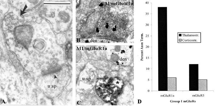
Figure 8.
Presynaptic mGluR1a in putative glutamatergic terminals from the thalamus and cerebral cortex. A, Shown is a mGluR1a-containing terminal forming an asymmetric axospinous synapse (arrowhead). The peroxidase deposit is indicated by a large arrow. B, C, Depicted are thalamostriatal (B) and corticostriatal (C) BDA-labeled boutons (asterisks in B and C) that express mGluR1a immunoreactivity (gold labeling) in the postcommissural putamen. In these micrographs, BDA has been revealed with DAB, whereas mGluR1a immunoreactivity is localized with gold particles. u. sp, Unlabeled spine; den, dendrite. Scale bars: (in A) B, 0.5 μm; C, 0.8 μm. D, Histogram that compares the relative abundance of thalamostriatal and corticostriatal terminals that express mGluR1a immunoreactivity in the monkey putamen. Note the preponderance of presynaptic mGluR1a labeling in thalamostriatal boutons. Term., Terminals; Thalamostr., thalamostriatal; Corticostr., corticostriatal.

