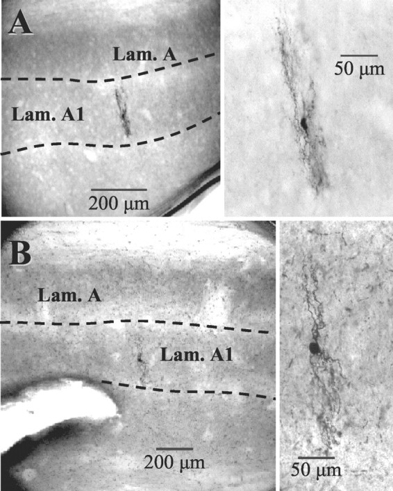
Figure 2.

Intralaminar interneurons from lateral geniculate nucleus. A, B, Left panels show location of two interneurons studied in the geniculate lamina. The dendritic trees of the cells were always perpendicular to the long axis of lamina A1 (Lam. A1). A, B, Right panels show higher magnification of interneurons. Note the small soma size and complicated dendritic architecture typical for this class of neurons.
