Figure 2.
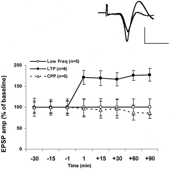
TBS induces LTP in the mPFC. This LTP is blocked by the NMDA receptor antagonist CPP. The increase in EPSP amplitude (LTP) was measured as a percentage of baseline value immediately before TBS to the BLA. Delivering TBS to the BLA induced a robust and long-lasting increase of the amplitude of the evoked field potential in the mPFC, reflecting the potentiation of the amygdala–PFC pathway. This group of LTP was significantly different from the low-frequency stimulation (Low Freq) controls at all the time points after TBS (F(5,7) = 5.149; p < 0.05). The level of potentiation in the LFS group was not significantly different from 100% at any time point. The injection of the competitive NMDA receptor antagonist (10 mg/kg) CPP 45 min before TBS significantly inhibited the induction of LTP; no increase of the EPSP amplitude was observed in the CPP-treated rats at any time point [t test for difference from baseline (100%); t(4) < 1; n = 5; NS]. Top left corner, Representative field potentials in the mPFC evoked during BLA stimulation immediately before and 90 min after TBS. The baseline (thin line) and the potentiated response are superimposed and are averages of 20 evoked responses each. Calibration: 0.2 mV, 10 msec.

