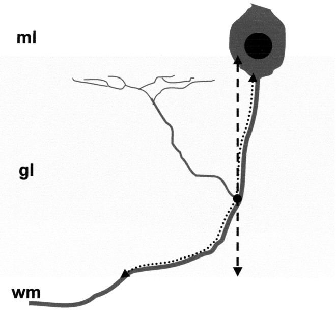
Figure 1.

Evaluation of the position of branching points along Purkinje axons. We examined Purkinje axons whose course from the Purkinje cell body to the white matter (wm) could be followed in a single section. The course of these axons through the granular layer (gl, shaded area) was reproduced (dotted arrow), and the position of the branch origin (black dot) was marked. In addition, to evaluate the position of branching points relative to the granular layer depth, the thickness of the granular layer was estimated along a line perpendicular to the cerebellar surface (dashed arrow), which passed through the branch origin. ml, Molecular layer.
