
Figure 2.
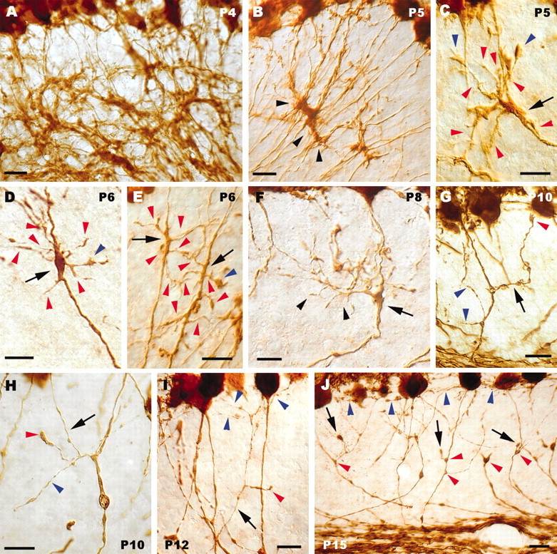
Sprouting, growth, and pruning of Purkinje axon collateral branches. A, Dense meshwork formed by calbindin-immunolabeled Purkinje axons in the nascent granular layer during the very first postnatal days. Arrowheads in B point to a clump of Purkinje axon swellings in the granular layer. C–E, Thickened or swollen segments (arrows) of individual Purkinje axons, from which numerous processes sprout (red arrowheads). Blue arrowheads indicate tiny growth cones at the tip of some of such newly formed processes. In F, a thick collateral branch (arrowheads) emanates from a focal enlargement (arrow) along the parent axon and spreads in the granular layer, giving off several second-order ramifications. The arrow in G indicates another collateral branch ending with a small growth cone (red arrowhead) in the vicinity of the Purkinje cell layer; two other ramifications, issued by the same corticofugal axon, are indicated by blue arrowheads. H, Thin collateral branch (arrow) ascending through the granular layer; a secondary ramification of this process ends with a large varicosity (red arrowhead). The blue arrowhead points to another process from the same intracortical plexus that ends with a small bouton. The thin branch shown in / (arrow) courses through the granular layer and terminates with some fine varicose chains (blue arrowheads) confined to the vicinity of the Purkinje cell layer; deeper in the granular layer, a shorter process belonging to the same plexus bears a terminal club (red arrowhead). Several presumptive pruning processes terminating with round clubs are indicated by arrows in J. Also note that the enlarged segments along the corticofugal neurites have disappeared, leaving small triangular varicosities (red arrowheads), whereas the fine terminal branches (some indicated by blue arrowheads) are confined to the most superficial portion of the granular layer. Anti-calbindin immunolabeling is shown in all panels. Postnatal ages are indicated on the micrographs. Scale bars: A, B, G, J, 20 μm; C–F, H, I, 10 μm.