
Figure 7.
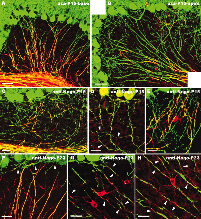
Morphology of Purkinje axon intracortical plexus after administration of 5′-azacytidine (A, B) or application of anti-Nogo-A antibodies (C–H). In the basal regions of cerebellar lobules from animals treated with 5′-azacytidine (A), myelinogenesis (as shown by MBP staining, red) is almost normal, and Purkinje axons (green) show the mature distribution pattern. In contrast, in apical regions of the same lobules (B), few MBP-positive processes are present in the granular layer, which is covered by numerous Purkinje axon profiles. C–E, Cerebellar regions close to the anti-Nogo-A injection site in animals killed 5 d after antibody injection (P15). Immunolabeling for MBP (C, red), Nogo-A (D, red; note the moderately stained Purkinje cell bodies), or CNP (E, red) shows sparse oligodendroglial profiles in the granular layer, where numerous Purkinje axon branches (green) remain (arrowheads in D point to collateral ramifications budding from a corticofugal axon). Numerous neuritic processes are also present in the vicinity of a CNP-positive oligodendrocyte in E. In rats killed 13 d after anti-Nogo-A injection (P23; F–H), Purkinje neurites are covered by MBP-stained oligodendroglial processes (F), whereas unmyelinated terminal branches (arrowheads) are confined within the upper granular layer. Anti-Nogo-A labeling of these cerebella (G, H) shows strongly stained oligodendrocytes, whose processes (arrowheads) twine around Purkinje neurites. Confocal images are from double-labeled sections for calbindin (green) and MBP (A–C, F, red), Nogo-A (D, G, H, red), and CNP (E, red). Scale bars: F–H, 20 μm; C–E, 25 μm; A, B, 40 μm.