
Figure 3.
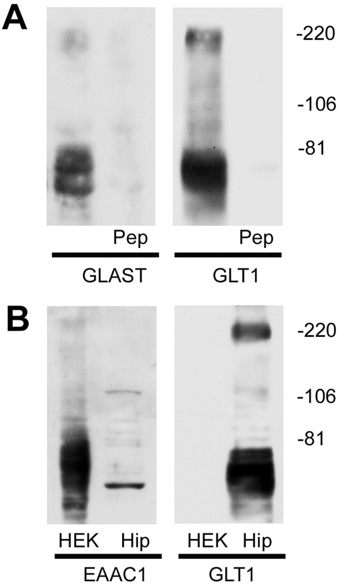
Immunoblot characterization of glutamate transporters antibodies. A, Protein samples from the hippocampus of adult rat were analyzed by immunoblot analysis using anti-GLAST (left) and anti-GLT1 (right), respectively. Both of the antibodies recognized bands at ∼66 and ∼220 kDa. Immunoreactivity was completely abolished when each antibody was preabsorbed with 90 μm of the peptide antigen (Pep), respectively. Control experiments were done with heterologous peptides (anti-GLT1 antibody preabsorbed with GLAST peptide and anti-GLAST antibody preabsorbed with GLT1 peptide), which had no effect on the immunoreactivity in hippocampal sample immunoblots. B, Immunoblot analysis showed that the anti-GLT1 antibody could not recognize EAAC1 protein. Protein samples from HEK 293 cells expressing EAAC1 protein (HEK) and hippocampus (Hip) were subjected to SDS-PAGE and immunoblotted with the antibodies of EAAC1 and GLT1, respectively. Antibody to EAAC1 recognized bands in EAAC1 cDNA-transfected HEK 293 cells as well as in rat hippocampal tissue (left), whereas GLT1 antibody only recognized the bands in rat hippocampal tissue (right). The immunoblots were representative of three independent experiments.