
Figure 1.
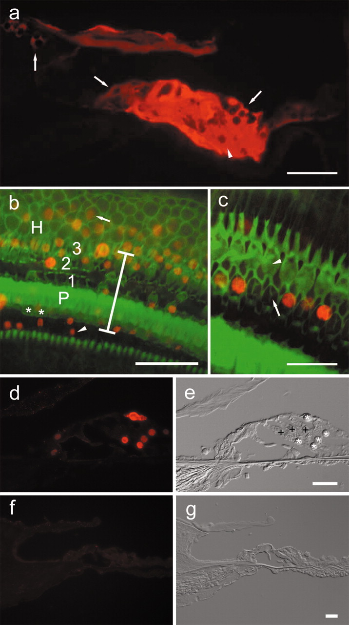
Epifluorescence ofβ-galactosidase and Math1 in the cochlear epithelium 4 d after Ad.Math1.11D inoculation. a, A cryosection showing β-galactosidase immunoreactivity in interdental(left arrow), inner sulcus(middle arrow), Hensen (right arrow), and supporting cells of the organ of Corti (arrowhead) in a cryosection of the third cochlear turn. b, A whole mount showing that Math1-positive nuclei (red) are in the inner sulcus (arrowhead), organ of Corti (vertical bar spans organ of Corti area; asterisk depicts inner hair cells; P depicts pillar cells; 1, 2, and 3 are first, second, and third row outer hair cells, respectively), and in the Hensen cell area (H) in which Math1-positive nuclei (arrow) are observed >30 μm outside the organ of Corti. Phalloidin staining (green) identifies surviving hair cells and sites of hair cell loss. c, Remaining hair cells adjacent to the inoculation site (phalloidin stain, green) are Math1 negative (arrowhead points to third row, outer hair cells). Some of the nonsensory cells that replaced lost hair cells (arrow in second row, outer hair cell area) are Math1 positive (red). d, e, Cryosection (d) of second turn of Ad.Math1.11D-inoculated organ of Corti showing Math1 immunoreactivity in nuclei of nonsensory cells (*). Outer hair cells (+) and several other cell types are negative. Nomarski optics image of same cryosection (e) identified cells shown in d. f, g, Cryosection of second-turn auditory epithelium of Ad.LacZ-inoculated cochlea. Math1 immunoreactivity is negative (f). Nomarski optics image of same cryosection (g) identifies cells. Scale bars: a, b, 50 μm; c, d–g, 25 μm.