
Figure 3.
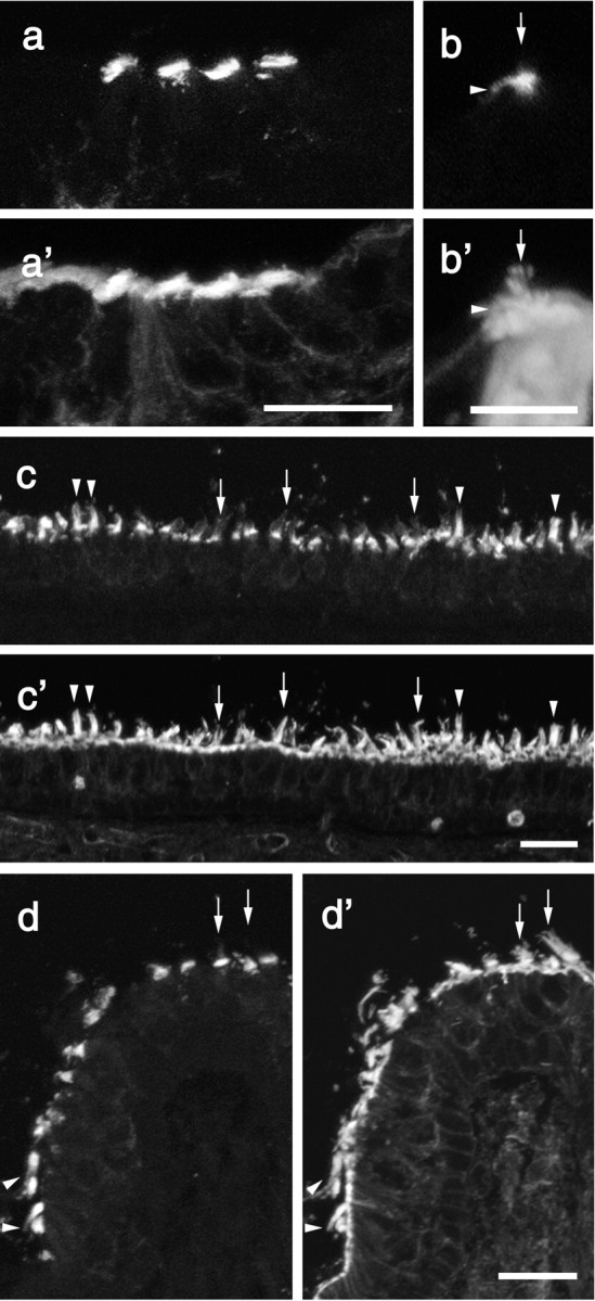
Fluorescence micrographs from mouse inner ear sections double labeled with affinity-purified antibodies to Ptprq (a-d) and rhodamine phalloidin (a′-d′).On cochlear inner hair cells (b,b′,arrows)and hair cells in the striolar regions of the utricular macula (c,c′,arrows) and at the apex of the crista (d, d′, arrows), anti-Ptprq staining is concentrated at the proximal end of the hair bundle. With hair bundles in the extrastriolar regions of the utricular macula (c, c′, arrowheads) and peripheral region of the crista (d, d′, arrowheads), the hair bundle is stained up to its tip. Sections are from mice at P2 (a), P21 (b, c), and P15 (d). Scale bars: a, 20 μm; b, 10 μm; c, d, 20 μm.