
Figure 8.
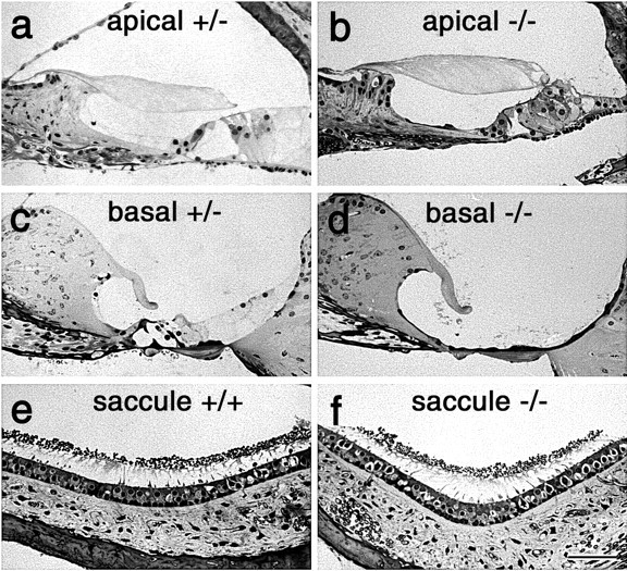
Structure of cochlea in wild-type, Ptprq-TM-KO, and Ptprq-CAT KO mice. Light micrographs of 1-μm-thick, toluidine blue-stained sections from the apical (a, b) and basal (c, d) regions of the cochlea, and the saccule(e,f),ofheterozygous(a,c),wild-type(e),andhomozygous(b, d,f)3-month-oldPtprq-CAT-KO mice. Note the complete loss of the organ of Corti in the basal end of the cochlea in the homozygous Ptprq-CAT-KO (d) mouse. Scale bar, 50 μm.