
Figure 9.
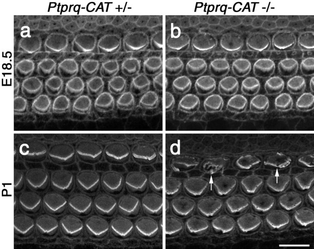
Phalloidin staining of hair bundles in Ptprq-CAT-KO mice. Confocal micrographs of the basal end of phalloidin-stained cochlear whole mounts from heterozygous (a, c) and homozygous Ptprq-CAT-KO(b,d)mice at E18.5(a,b)and P1(c,d). Defects in hair-bundle structure are first apparent in the inner hair cells (d, arrows) at P1. Scale bar, 10 μm.