
Figure 2.
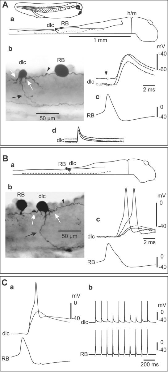
Paired recordings show that RB sensory neurons strongly excite sensory pathway dlc interneurons at a short and constant latency. A, a, Tadpole 5-6 mm long and a scale diagram of the CNS show a filled RB and dlc interneuron. The dlc axon (dotted line) is on the opposite left side; the hindbrain to midbrain border is indicated (h/m). b, RB and dlc interneuron at the cut dorsal edge of the spinal cord in the fifth segment on the right side. The spherical RB soma has an ascending axon and descending axon (arrowhead) with possible contact sites onto dlc dendrites (white arrows). The multipolar dlc soma has oblique dendrites and a ventral initial process leading to a ventral commissural axon (black arrow). c, Current injection evoking an action potential in RB leads to a large EPSP in the dlc interneuron at short latency (three traces overlapped; the whole EPSP, trace length, 100 msec, is shown in d). The RB impulse causes a small artifact in the dlc interneuron (arrowhead). B, a, In this case, the RB is caudal to the dlc interneuron. b, Ascending RB axon (arrowhead) with possible contacts to the dlc interneuron (white arrows) that has a commissural axon (black arrow). c, Current-evoked RB action potentials lead to variable amplitude dlc interneuron EPSPs, two of which reach a threshold for a dlc action potential. C, a, An example in which current-evoked action potentials in an RB in the sixth segment lead to large, reliable, constant-latency EPSPs in a dlc interneuron. Even with -30 pA hyperpolarizing current injected into the dlc interneuron, these EPSPs can reach threshold and evoke dlc interneuron action potentials (time scale as in B). The reliability of the EPSP is shown by repeated current pulses to the RB neuron.