
Figure 6.
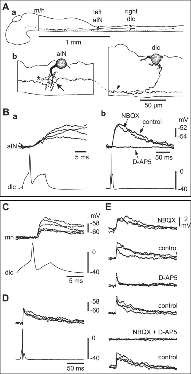
Paired recordings show that sensory pathway dlc interneurons excite contralateral neurons by activating glutamate receptors. A, a, Scale drawings of the CNS viewed from the left side to show a dlc interneuron on the right side of the spinal cord with a commissural axon that branches on the left side, where its ascending axon (dotted) has possible contact sites onto an aIN. b, The multipolar dlc interneuron soma has an ascending axon (arrowhead) with possible contact points (arrow) on dendrites of the aIN, the axon (asterisk) of which has been omitted for clarity.B,a,Current injection evoking an action potential in the dlc interneuron shown in Aleads to slow-rise, small EPSPs in the aIN at short latency(four over lapped traces).b,The same neuron pair shows that the long-duration EPSP is blocked by 50 μm d-AP-5 but is little affected by 5 μm NBQX. C, Small, short-latency EPSPs (six overlapped traces) in a mn produced by current-evoked dlc interneuron impulses (dlc). D, The same recording as C showing the long duration of the EPSP. E, Effects of glutamate receptor antagonists on EPSPs evoked in the motoneuron by dlc interneuron action potentials. The long, slow component of the EPSP is blocked by 50 μm d-AP-5 and recovers in wash. The fast initial component is blocked by 5 μm NBQX. After recovery in wash, the EPSP is blocked by simultaneous application of 5 μm NBQX and 50 μm d-AP-5 and returns after wash. Antagonists were drop-applied (three overlapped traces; time scale as in D).