
Figure 7.
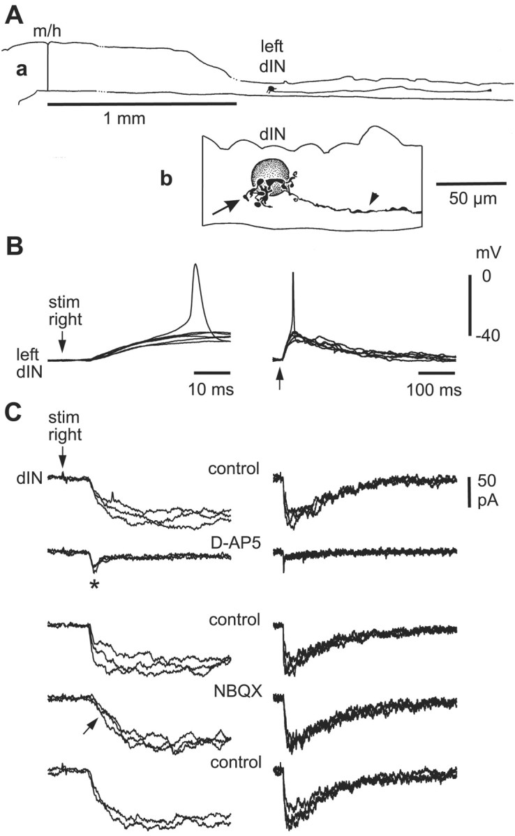
Pharmacology of responses to contralateral skin stimulation in a dIN. A, a, Scale drawing of the CNS viewed from the left side to show the dIN on the left side of the spinal cord with a descending axon. b, Details of the dIN multipolar soma with short dendrites (arrow) and a descending axon (arrowhead). B, Contralateral tail-skin stimulation (arrow) produces a slow-rise EPSP at constant latency that, in one trace, evokes a spike in the dIN. C, Pharmacology of EPSCs in the same dIN at a holding potential of -65 mV. d-AP-5 blocks a slow component to reveal a small fast EPSC (asterisk), which is blocked by NBQX (arrow). Antagonists were microperfused, and the circulating saline contained 10 μm mecamylamine and 2 μm strychnine to block any cholinergic and glycinergic currents (three overlapped traces; time scales as in B).