Figure 5.
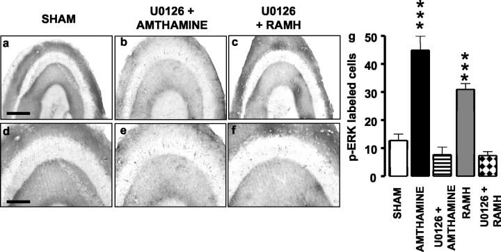
Influence of U0126 on amthamine- or RAMH-elicited increase of p-ERK immunoreactivity in hippocampal slices. a-f, Representative photomicrographs for p-ERK immunoreactivity in hippocampal slices. Scale bar: a-c, 400 μm, d-f, 200 μm. g, Summary histograms show the number of neurons staining positive for p-ERK in controls (n = 16), in slices treated with 0.1 μm amthamine alone (n = 10) or associated with 2 μm U0126 (n = 3), and 0.1 μm RAMH alone (n = 10) or associated with 2 μm U0126 (n = 3). Shown are means ± SEM. ***p< 0.001 versus control (ANOVA and Newman-Keuls multiple comparison test).

