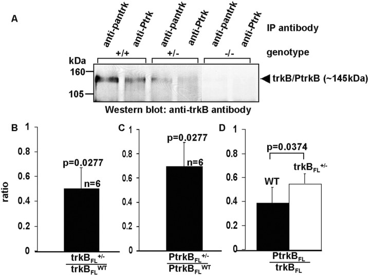
Figure 4.
A, Immunoprecipitation-immunoblot analysis of trkB phosphorylation (activation). Lysates were obtained from the retinas of WT (+/+), trkBFL+/-, and trkBFL-/- mice, immunoprecipitated with anti-pan-trk or anti-Ptrk, and probed with an antibody against the extracellular domain of trkB. B, Ratio of retinal trkBFL in trkBFL+/- mice to retinal trkBFL in WT mice. C, Ratio of retinal PtrkBFL in trkBFL+/- mice to retinal PtrkBFL in WT mice. D, Retinal PtrkBFL/trkBFL in WT and trkBFL+/- mice. p values indicate significance of intergroup differences.

