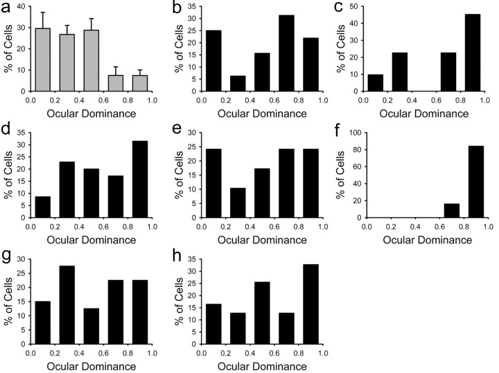
Figure 3.
Reduced ocular dominance plasticity in animals exposed to alcohol from P10-P30. a, The histogram with error bars pools together results from alcohol-treated animals that received normal binocular vision. The primary visual cortex of these animals was characterized by a large number of binocular neurons with a predominance of the contralateral eye, as occurs in normal animals. b-h, After 3 d of monocular deprivation, the histograms for individual animals treated with alcohol show a reduced ocular dominance shift (b-g) with a single exception (f). In these animals, most neurons could still be driven by the deprived contralateral eye.

