
Figure 2.
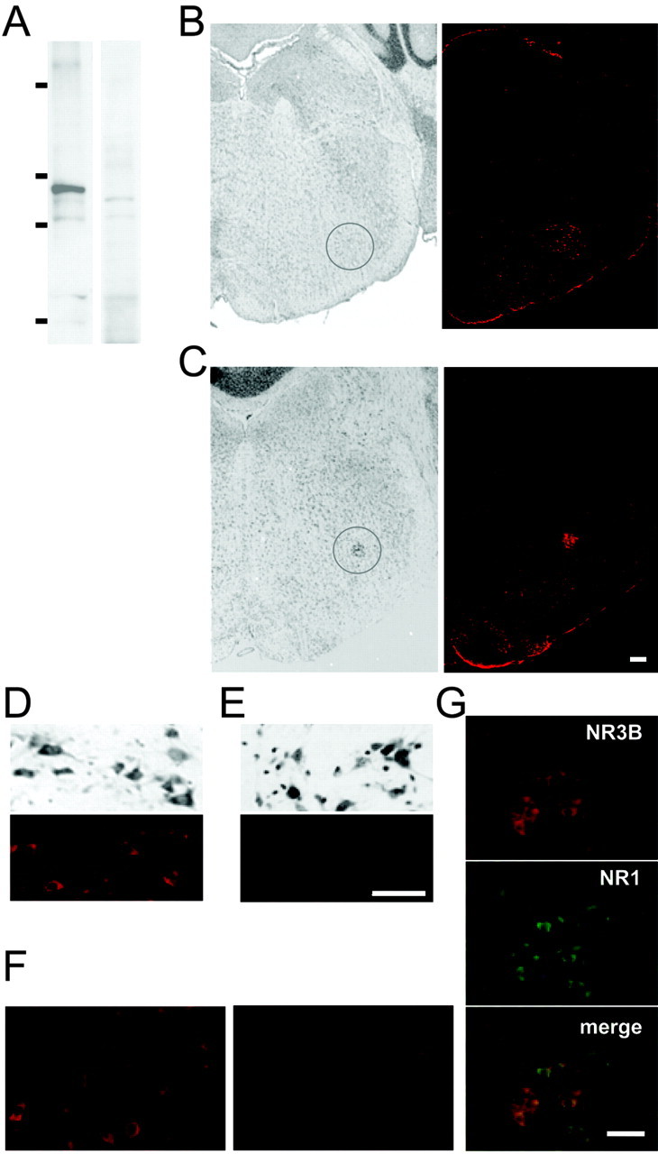
NR3B immunoreactivity in motor neurons in the brainstem. A, Immunoblot analysis. Lysates of mice whole brain were subjected to immunoblot analysis using anti-NR3B antibody (left panel) or anti-NR3B antibody preabsorbed with the peptide used to immunize the rabbit (right panel). Molecular weight markers correspond to 203, 120, 90, and 51 kDa. B, C, Coronal sections of adult mouse brainstem were stained with cresyl violet (left panels), and adjacent sections were stained with anti-NR3B antibody and visualized by Alexa 546-conjugated secondary antibody (right panels). The circled region corresponds to the facial nucleus (B) or the ambiguous nucleus (C). D, E, Enlarged view of the facial nucleus (D) and the abducent nucleus (E) in the same section. Top panels, Corresponding cresyl violet staining. F, Before (left panel) and after (right panel) absorption of anti-NR3B antibody with the peptide. These fields correspond to the facial nucleus. G, Double immunostaining of NR3B (top panel) and NR1 (middle panel) in the ambiguous nucleus. Fluorescent images were overlaid (bottom panel) to show the colocalization. Scale bars: C (also applies to B, C), E, (also applies to D-F), G, 100 μm.