
Figure 5.
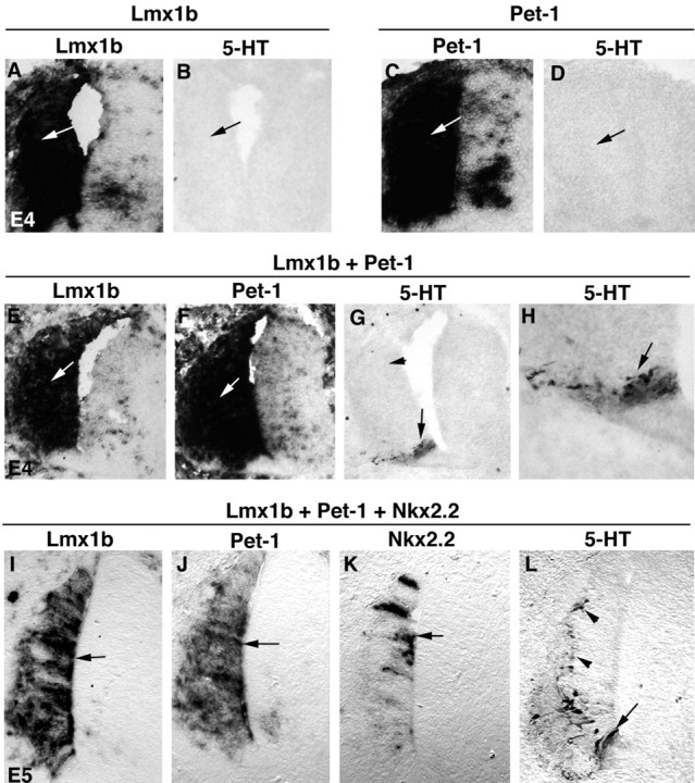
Lmx1b, Pet-1, and Nkx2.2 in combination are sufficient to induce 5-HT cell fate in the chick spinal cord. Transverse sections through the spinal cord are shown. Plasmids were electroporated at E2 and analyzed at E4 (A-H) or E5 (I-L). The plasmids used for electroporation are shown above the lines. Expression of the transgenes (the names of which are shown at the top of the panels and below the lines) was detected by in situ hybridization. Also, 5-HT immunostaining was performed (B, D, G, H, L). H is the high magnification of the positive area shown in G.